. 2019 Mar;52(2):e12557.
doi: 10.1111/cpr.12557. Epub 2018 Nov 28.

Matrix stiffness regulates arteriovenous differentiation of endothelial progenitor cells during vasculogenesis in nude mice

Affiliations

Matrix stiffness regulates arteriovenous differentiation of endothelial progenitor cells during vasculogenesis in nude mice

Changyue Xue et al. Cell Prolif. 2019 Mar.

Abstract

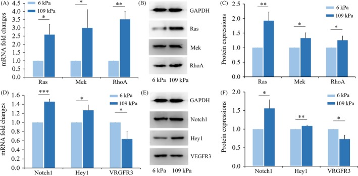
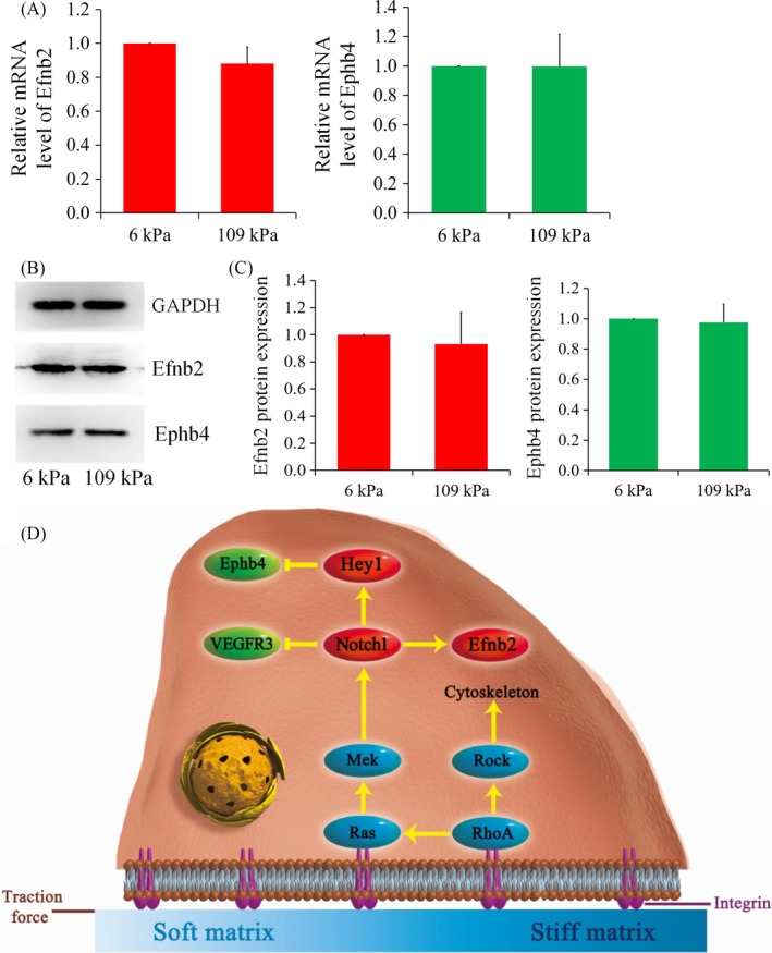
Objectives: The aim of the study was to investigate the effect of matrix stiffness on arteriovenous differentiation of endothelial progenitor cells (EPCs) during vasculogenesis in nude mice.

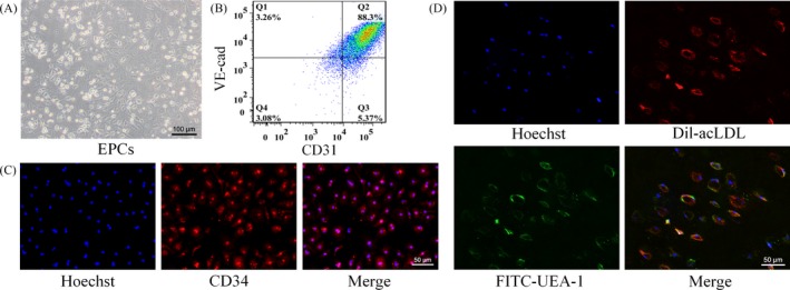
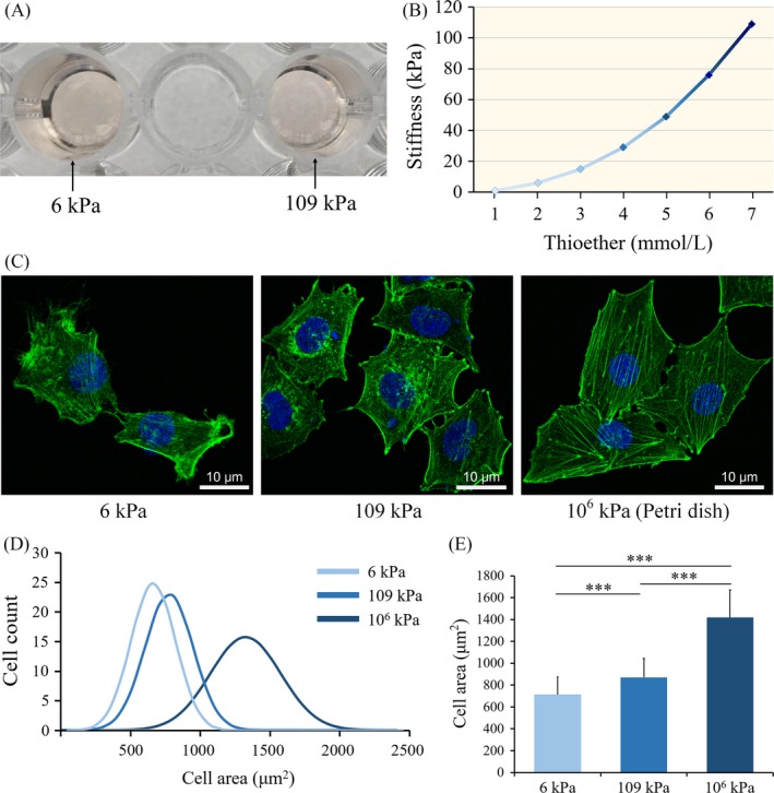
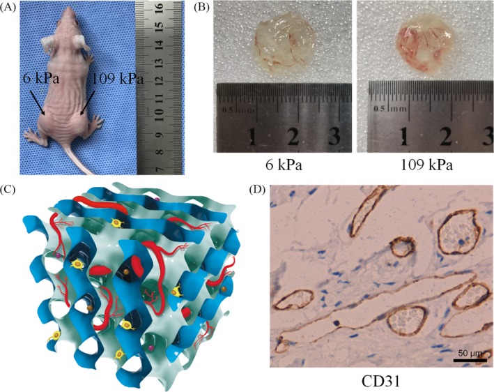
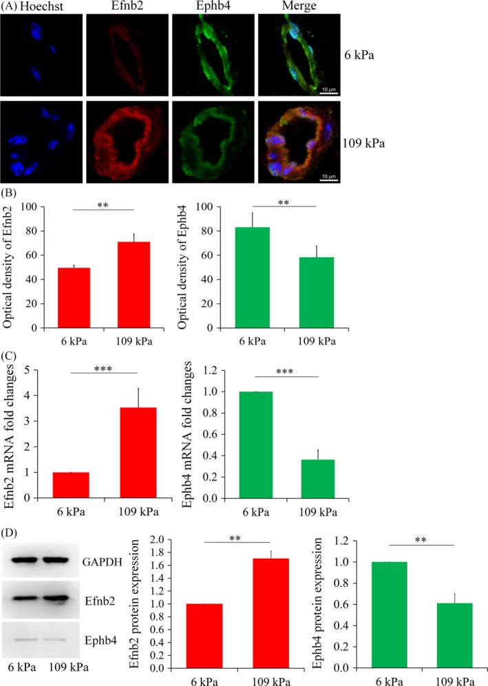
Materials and methods: Dextran hydrogels of differing stiffnesses were first prepared by controlling the crosslinking reaction to generate different thioether bonds. Hydrogels with stiffnesses matching those of the arterial extracellular matrix and venous extracellular matrix were separately combined with mouse bone marrow-derived EPCs and subcutaneously implanted on either side of the backs of nude mice. After 14 days, artery-specific marker Efnb2 and vein-specific marker Ephb4 in the neovasculature were detected to determine the effect of matrix stiffness on the arteriovenous differentiation of EPCs in vivo.

Results: Fourteen days after the implantation of the EPC-loaded dextran hydrogels, new blood vessels were observed in both types of hydrogels. We further verified that matrix stiffness regulated the arteriovenous differentiation of EPCs during vasculogenesis via the Ras/Mek pathway.

Conclusions: Matrix stiffness regulates the arteriovenous differentiation of EPCs during vasculogenesis in nude mice through the Ras/Mek pathway.

Keywords: Ras/Mek pathway; arteriovenous development; matrix characteristics; tissue engineering; vasculogenesis.


Conflict of interest statement

The authors declare that they have no competing interests.

Figures

Figure 1
Figure 1
Characterization of EPCs. A, Image of the EPCs observed under an inverted light microscope; B, Flow cytometry for CD31 and VE‐cad expression of EPCs. Percentage of CD31+/VE‐cad+ cells among total EPCs is indicated (Q2); C, Positive staining for the stem/progenitor cell marker CD34; D, Ulex europaeus agglutinin‐1 binding (green) and 1,1‐dioctadecyl‐3,3,3,3‐tetramethylindo‐carbocyanin‐eperchlorate‐labelled acetylated low‐density lipoprotein uptake (red) in EPCs
Figure 2
Figure 2
A, Two dextran hydrogels with different stiffnesses after crosslinking; B, Stiffnesses of dextran hydrogels with different dextran (thioether bond) contents; five different samples were tested as indicated; C, Fluorescent images of the cytoskeleton (phalloidin staining of F‐actin) of EPCs on matrix of the indicated stiffness. Scale bars are 20 μm; D, Distributions of cell area of EPCs on matrices of varying degrees of stiffness; E, Quantitative assessment of cell morphology; 100 cells were measured in each group. *P < 0.05, **P < 0.01, ***P < 0.001. Data shown are representative of three independent experiments
Figure 3
Figure 3
A, Injection of the hydrogel‐EPC composites on either side of the backs of nude mice; B, Vasculogenesis status after 14 d of hydrogel implantation; C, Schematic representation of the vasculogenesis model in the hydrogel support material. Brown and purple balls represent vascular growth factors; D, Immunohistochemical staining of CD31 in hydrogel (6 kPa) slices. Scale bar is 50 μm
Figure 4
Figure 4
A, Double immunofluorescence staining of arteriovenous marker proteins (scale, 10 μm) in hydrogels with different stiffnesses; B, Statistical analysis of the fluorescence intensities. Each experimental value is expressed as the mean ± standard deviation; C, Gene transcript levels of the arteriovenous markers in the newly formed vessels in the hydrogels of different stiffnesses. All groups of genes were first normalized to internal references, then normalized to the control group (6 kPa group); D, Western blotting for and statistical analysis of Efnb2 and Ephb4 expression in the newly formed vessels in the hydrogels of different stiffnesses. Data shown were representative of three independent experiments; *P < 0.05, ** P < 0.01, ***P < 0.001
Figure 5
Figure 5
Small GTP‐binding protein pathways and Notch signal transduction are activated by matrix with high stiffness. A, Real‐time PCR analyses of Ras, MAP kinase‐ERK kinase (Mek) and Ras homologue family member A (RhoA) expression; B, Western blot analyses showing protein expression of Ras, Mek and RhoA; C, The protein expressions of Ras, Mek and RhoA were quantitated, and data are shown as a histogram. Each experimental value is expressed as the mean ± standard deviation; D, Real‐time PCR analyses of Notch1, hairy/enhancer‐of‐split related with YRPW motif 1(Hey1) and vascular endothelial growth factor receptor 3 (VEGFR3) expression; E, Western blotting of Notch1, Hey1 and VEGFR3; F, Protein expressions of Notch1, Hey1 and VEGFR3 were quantitated and data are shown as a histogram. *P < 0.05, **P < 0.01, ***P < 0.001. Data shown are representative of three independent experiments
Figure 6
Figure 6
Results of inhibitor study. A, The mRNA levels of Efnb2 and Ephb4 both showed no difference between 6 kPa group and 109 kPa group different after treatment with farnesylthiosalicylic acid; B, C, Western blot analyses also showed that the regulatory role of matrix stiffness was blocked after treatment with farnesylthiosalicylic acid. Data shown are representative of three independent experiments; D, Schematic diagram illustrating the mechanism by which the arteriovenous differentiation of EPCs during vasculogenesis is regulated in response to matrix stiffness

Similar articles

Cited by

References

    1. Augustin HG, Koh GY. Organotypic vasculature: from descriptive heterogeneity to functional pathophysiology. Science. 2017;357(6353):eaal2379. - PubMed
    1. Bompais H, Chagraoui J, Canron X, et al. Human endothelial cells derived from circulating progenitors display specific functional properties compared with mature vessel wall endothelial cells. Blood. 2004;103(7):2577. - PubMed
    1. Asahara T, Masuda H, Takahashi T, et al. Bone marrow origin of endothelial progenitor cells responsible for postnatal vasculogenesis in physiological and pathological neovascularization. Circ Res. 1999;85(3):221‐228. - PubMed
    1. Hur J, Yoon CH, Kim HS, et al. Characterization of two types of endothelial progenitor cells and their different contributions to neovasculogenesis. Arterioscler Thromb Vasc Biol. 2004;24(2):288‐293. - PubMed
    1. Obi S, Yamamoto K, Shimizu N, et al. Fluid shear stress induces arterial differentiation of endothelial progenitor cells. J Appl Physiol. 2009;106(1):203‐211. - PubMed