

Endothelial colony-forming cell therapy for heart morphological changes after neonatal high oxygen exposure in rats, a model of complications of prematurity
- PMID: 30485704
- PMCID: PMC6260919
- DOI: 10.14814/phy2.13922
Endothelial colony-forming cell therapy for heart morphological changes after neonatal high oxygen exposure in rats, a model of complications of prematurity
Abstract
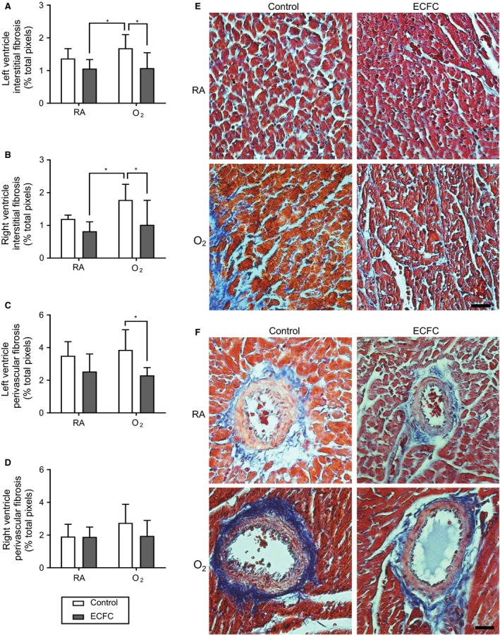
Very preterm birth is associated with increased cardiovascular diseases and changes in myocardial structure. The current study aimed to investigate the impact of endothelial colony-forming cell (ECFC) treatment on heart morphological changes in the experimental model of neonatal high oxygen (O2 )-induced cardiomyopathy, mimicking prematurity-related conditions. Sprague-Dawley rat pups exposed to 95% O2 or room air (RA) from day 4 (P4) to day 14 (P14) were randomized to receive (jugular vein) exogenous human cord blood ECFC or vehicle at P14 (n = 5 RA-vehicle, n = 8 RA-ECFC, n = 8 O2 -vehicle and n = 7 O2 -ECFC) and the hearts collected at P28. Body and heart weights and heart to body weight ratio did not differ between groups. ECFC treatment prevented the increase in cardiomyocyte surface area in both the left (LV) and right (RV) ventricles of the O2 group (O2 -ECFC vs. O2 -vehicle LV: 121 ± 13 vs. 179 ± 21 μm2 , RV: 118 ± 12 vs. 169 ± 21 μm2 ). In O2 rats, ECFC treatment was also associated with a significant reduction in interstitial fibrosis in both ventricles (O2 -ECFC vs. O2 -vehicle LV: 1.07 ± 0.47 vs. 1.68 ± 0.41% of surface area, RV: 1.01 ± 0.74 vs. 1.77 ± 0.67%) and in perivascular fibrosis in the LV (2.29 ± 0.47 vs. 3.85 ± 1.23%) but in not the RV (1.95 ± 0.95 vs. 2.74 ± 1.14), and with increased expression of angiogenesis marker CD31. ECFC treatment had no effect on cardiomyocyte surface area or on tissue fibrosis of RA rats. Human cord blood ECFC treatment prevented cardiomyocyte hypertrophy and myocardial and perivascular fibrosis observed after neonatal high O2 exposure. ECFC could constitute a new regenerative therapy against cardiac sequelae caused by deleterious conditions of prematurity.
Keywords: Cell therapy; heart; oxygen-induced cardiomyopathy; preterm birth.
© 2018 The Authors. Physiological Reports published by Wiley Periodicals, Inc. on behalf of The Physiological Society and the American Physiological Society.
Figures


References
-
- Asahara, T. , Kawamoto A., and Masuda H.. 2011. Concise review: circulating endothelial progenitor cells for vascular medicine. Stem Cells 29:1650–1655. - PubMed
-
- Bensley, J. G. , Stacy V. K., De Matteo R., Harding R., and Black M. J.. 2010. Cardiac remodelling as a result of pre‐term birth: implications for future cardiovascular disease. Eur. Heart J. 31:2058–2066. - PubMed
MeSH terms
Substances
Grants and funding
LinkOut - more resources
Full Text Sources
Medical
Research Materials
