Review
. 2019 Feb 10:81:375-402.
doi: 10.1146/annurev-physiol-020518-114320. Epub 2018 Nov 28.

Cell Death in the Lung: The Apoptosis-Necroptosis Axis

Affiliations
Review

Cell Death in the Lung: The Apoptosis-Necroptosis Axis

Maor Sauler et al. Annu Rev Physiol. .

Abstract

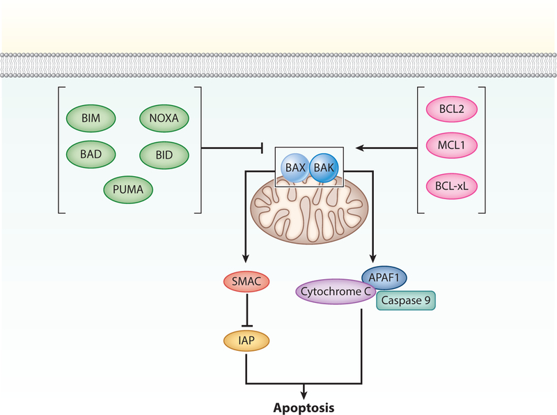
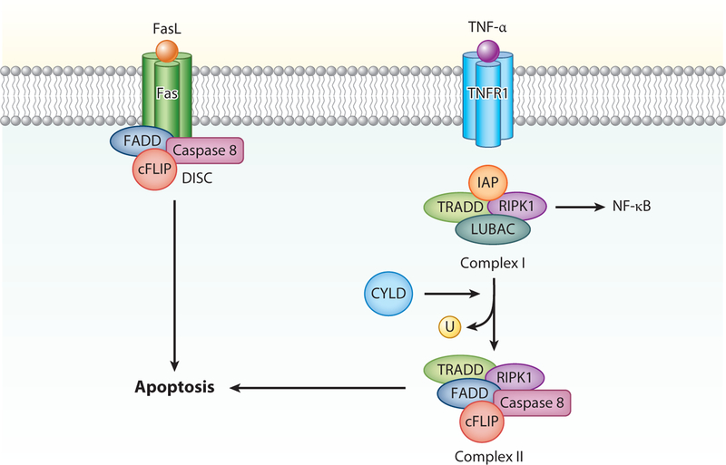
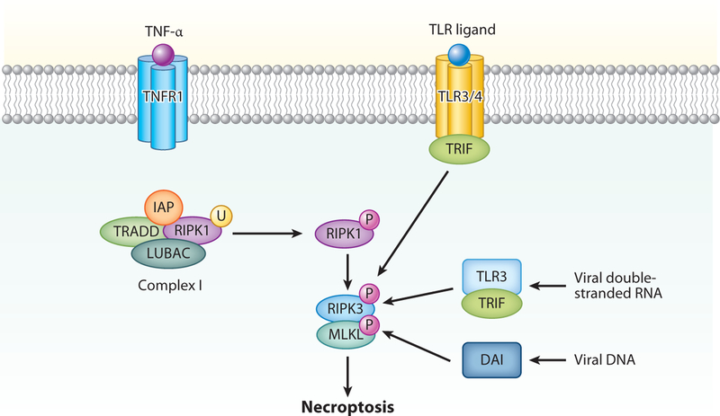
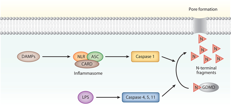
Regulated cell death is a major mechanism to eliminate damaged, infected, or superfluous cells. Previously, apoptosis was thought to be the only regulated cell death mechanism; however, new modalities of caspase-independent regulated cell death have been identified, including necroptosis, pyroptosis, and autophagic cell death. As an understanding of the cellular mechanisms that mediate regulated cell death continues to grow, there is increasing evidence that these pathways are implicated in the pathogenesis of many pulmonary disorders. This review summarizes our understanding of regulated cell death as it pertains to the pathogenesis of chronic obstructive pulmonary disease, asthma, idiopathic pulmonary fibrosis, acute respiratory distress syndrome, and pulmonary arterial hypertension.

Keywords: acute respiratory distress syndrome; apoptosis; asthma; chronic obstructive pulmonary disease; idiopathic pulmonary fibrosis; necroptosis; pulmonary arterial hypertension.


Figures

Figure 1
Figure 1
Intrinsic apoptosis. The step that commits cells to undergo apoptosis is permeabilization of the mitochondrial outer membrane (MOMP). MOMP occurs when B-cell lymphoma 2 (BCL2)-associated X apoptosis regulator (BAX) and BCL2 antagonist/killer 1 (BAK) form outer mitochondrial membrane pores. This process is promoted by BH3-only proteins, including BCL2-associated death promoter (BAD), p53-upregulated binding component (PUMA), BCL2-like 11 (BIM), phorbol-12-myristate-13-acetate-induced protein (NOXA), and BH3-interacting domain death agonist (BID); and antagonized by antiapoptotic BCL2 proteins, including BCL2, B-cell lymphoma extra large (BCL-xL), and myeloid leukemia cell differentiation protein (MCL1). MOMP causes the release of cytochrome c and second mitochondria–derived activator of caspase (SMAC). Cytochrome c binds apoptotic protease activating factor 1 (APAF1) and initiator caspase 9 to form the apoptosome, where caspase 9 is activated. SMAC neutralizes the cytoplasmic proteins maintained by cells to restrain caspase activation (inhibitor of apoptosis proteins, IAPs).
Figure 2
Figure 2
Extrinsic apoptosis. Ligand binding to Fas results in the formation of the death-inducing signaling complex (DISC), composed of Fas-associated death domain (FADD), cellular FLICE-inhibitory protein (cFLIP), and caspase 8. Upon activation, cleavage of caspase 8 leads to cleavage of executioner caspases and apoptosis. The consequences of TNF receptor 1 (TNFR1) activation depends on posttranslational modifications of another protein recruited to Complex 1, receptor-interacting protein kinase 1 (RIPK1). Upon TNFR1-associated death domain (TRADD)-dependent recruitment, RIPK1 can be ubiquitinated by inhibitor of apoptosis proteins (IAPs) and linear ubiquitination chain assembly complex (LUBAC). Ubiquitinated RIPK1 (U) promotes inflammation and cell survival by activating protein kinase signaling and IκB kinase (IKK)-dependent NF-κB activation. If IAPs are absent or inhibited, RIPK1 is deubiquitinated by cylindromatosis (CYLD). Consequently, RIPK1 forms a complex with FADD and caspase 8 in the cytosol (Complex II). Similar to signaling by Fas, this complex is also regulated by cFLIP proteins and can lead to caspase 8 activation and apoptosis.
Figure 3
Figure 3
Necroptosis can occur as a consequence of TNF receptor 1 (TNFR1) activation. Similar to extrinsic apoptosis, RIPK1 ubiquitination is favored in the absence of inhibitor of apoptosis proteins (IAPs) and the presence of linear ubiquitination chain assembly complex (LUBAC). In the absence of caspase 8, RIPK1 is phosphorylated (P) and interacts with RIPK3, leading to oligomerization and phosphorylation of mixed lineage kinase domain-like pseudokinase (MLKL). DNA-dependent activator of interferon (DAI) regulatory factors can detect viral DNA and activate RIPK3-mediated necroptosis. Toll-like receptor-3 (TLR3) can also detect viral DNA and, via TLR-domain-containing adapter-inducing interferon-β (TRIF), can activate RIPK3 independent of RIPK1, also leading to necroptosis. Similarly, TLR4 can activate RIPK3 via TRIF independently of RIPK1, which also leads to necroptosis.
Figure 4
Figure 4
Pyroptosis. Pyroptosis is initiated when damage-associated molecular patterns (DAMPs) activate the NOD-like receptor (NLR) leading to the formation of the inflammasome protein complex, which includes caspase activation and recruitment domain (CARD) and apoptosis-associated speck protein containing a CARD (ASC). Inflammasome activation leads to cleavage of procaspase 1, which in turn cleaves the N terminus of gasdermin D (GDMD) to generate pore forming N-terminal fragments. These fragments create pores in the cell membrane that kill the cells. Pyroptosis can also be initiated when lipopolysaccharide (LPS) activates caspases 4, 5, and 11 directly, which also leads to GDMD cleavage and pyroptosis.
Figure 5
Figure 5
Autophagy. Autophagy occurs under conditions of nutrient deprivation or cellular stress. This cellular stress activates UNC-51-like kinase 1 (ULK) complex and Beclin-1, via molecular target of rapamycin (mTOR) and 5’ adenosine monophosphate-activated protein kinase (AMPK). When activated, a membrane forms that includes the autophagic protein light chain 3 (LC3). Elongation of the membrane results in the formation of an autophagosome which engulfs the cellular components to be degraded. The autophagosome fuses with a lysosome, forming the autolysosome, which ultimately degrades the cellular components.

Similar articles

Cited by

References

    1. Elmore S 2007. Apoptosis: a review of programmed cell death. Toxicol. Pathol 35:495–516 - PMC - PubMed
    1. Ashkenazi A, Salvesen G. 2014. Regulated cell death: signaling and mechanisms. Annu. Rev. Cell Dev. Biol 30:337–56 - PubMed
    1. Kerr JF, Wyllie AH, Currie AR. 1972. Apoptosis: a basic biological phenomenon with wide-ranging implications in tissue kinetics. Br. J. Cancer 26:239–57 - PMC - PubMed
    1. Galluzzi L, Vitale I, Aaronson SA, Abrams JM, Adam D, et al. 2018. Molecular mechanisms of cell death: recommendations of the Nomenclature Committee on Cell Death 2018. Cell Death Differ 25:486–541 - PMC - PubMed
    1. Fuchs Y, Steller H. 2011. Programmed cell death in animal development and disease. Cell 147:742–58 - PMC - PubMed

Publication types

MeSH terms