. 2018 Nov 27;25(9):2417-2430.e5.
doi: 10.1016/j.celrep.2018.10.097.

Esrp1-Regulated Splicing of Arhgef11 Isoforms Is Required for Epithelial Tight Junction Integrity

Affiliations

Esrp1-Regulated Splicing of Arhgef11 Isoforms Is Required for Epithelial Tight Junction Integrity

SungKyoung Lee et al. Cell Rep. .

Abstract

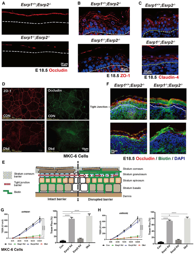
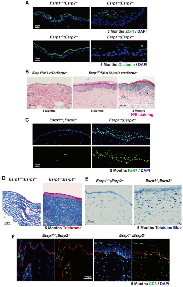
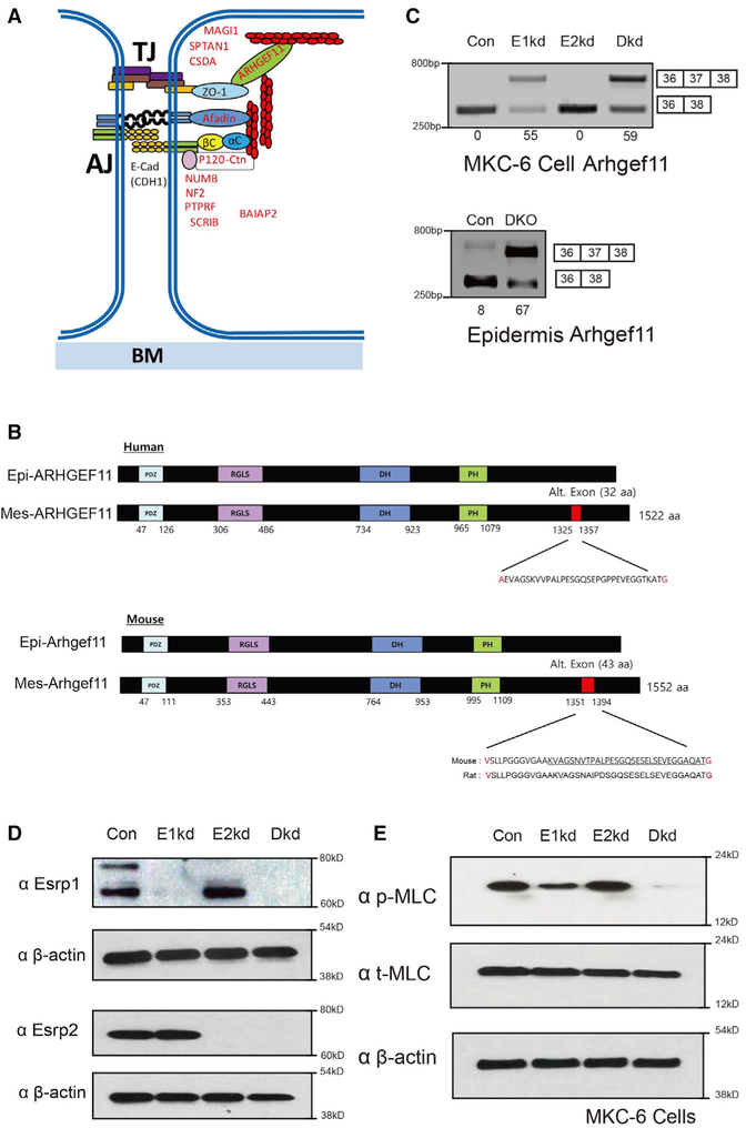
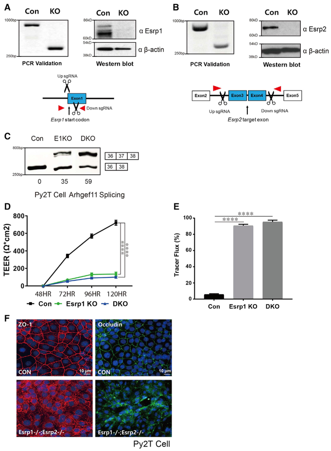
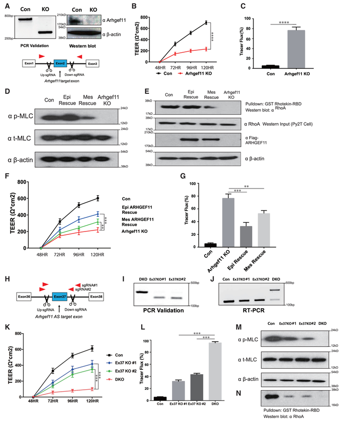
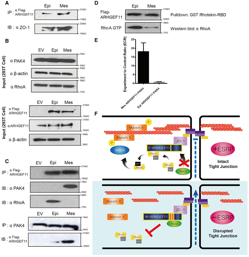
The epithelial-specific splicing regulators Esrp1 and Esrp2 are required for mammalian development, including establishment of epidermal barrier functions. However, the mechanisms by which Esrp ablation causes defects in epithelial barriers remain undefined. We determined that the ablation of Esrp1 and Esrp2 impairs epithelial tight junction (TJ) integrity through loss of the epithelial isoform of Rho GTP exchange factor Arhgef11. Arhgef11 is required for the maintenance of TJs via RhoA activation and myosin light chain (MLC) phosphorylation. Ablation or depletion of Esrp1/2 or Arhgef11 inhibits MLC phosphorylation and only the epithelial Arhgef11 isoform rescues MLC phosphorylation in Arhgef11 KO epithelial cells. Mesenchymal Arhgef11 transcripts contain a C-terminal exon that binds to PAK4 and inhibits RhoA activation byArhgef11. Deletion of the mesenchymal-specific Arhgef11 exon in Esrp1/2 KO epithelial cells using CRISPR/Cas9 restored TJ function, illustrating how splicing alterations can be mechanistically linked to disease phenotypes that result from impaired functions of splicing regulators.

Keywords: epidermis.


Figures

Figure 1.
Figure 1.. Esrp Ablation in Mouse Epidermis or Depletion in Keratinocytes Disrupts Tight Junctions and the Permeability Barrier
(A–C) Immunostaining for Occludin (A), ZO-1 (B), and Claudin-4 (C) in Esrp1−/−;Esrp2−/−DKO (double knockout) and control Esrp1+/+;Esrp2−/− epidermis showing disrupted Occludin continuity at the stratum granulosum (SG) layer. DAPI (blue). (C) Claudin-4 staining showing reduced thickness of DKO epidermis compared to the control. (D) Immunofluorescence localization of tight junction (TJ) markers in the mouse MKC-6 keratinocytes cell line 96 hr after a calcium shift. Analysis was done in cells transduced with a control non-targeting shRNA or shRNAs targeting Esrp1 and Esrp2. (E) Schematic representing the paracellular biotin permeability assay. (F) Control (Esrp1+/+;Esrp2−/−) or Esrp DKO (Esrp1−/−;Esrp2−/−) E18.5 mouse embryo skin injected intradermally with biotin was stained with streptavidin to follow the penetration of biotin (green) and counterstained with TJ marker Occludin (red) and DAPI (blue) to mark the TJs in the SG layer. (G and H) Transepithelial electrical resistance (TEER) was measured across confluent control, Esrp1, Esrp2, or Esrp1/2 shRNA knockdown MKC-6 keratinocyte cell monolayers stimulated with calcium for the indicated times suing Esrp1 shRNA#1 (G) and Esrp1 shRNA#2 (H). (G) and (H) used different Esrp1 shRNAs. Tracer flux of 4kDa FITC-dextran in control or Esrp depleted keratinocyte cells is indicated as percent tracer flux relative to a transwell with no cells. Error bars indicate means ± SDs, n = 3. Statistical significance comparing each group with control was determined by t test. *p < 0.05, **p < 0.005, ****p < 0.0001.
Figure 2.
Figure 2.. Postnatal Induction of Esrp Ablation in the Epidermis Leads to Disrupted TJ, Epidermal Thickening, Inflammation, and Scarring
(A) Control (Esrp1f/f; K5-rtTA;Esrp2−/−) or Esrp DKO (Esrp1f/f; K5-rtTA;tetO-Cre;Esrp2−/−) 5-month-old skin immunostained for ZO-1 and Occludin (green). (B) Longitudinal paraffin section from mice back skin of control and Esrp1f/f; K5-rtTA;tetO-Cre;Esrp2−/− mice. H&E stain reveals that Esrp ablation causes hyperthickening of the epidermis. (C) Ki-67 proliferation assay. Ki-67+cells (green) and DAPI (blue). (D) Masson’s trichrome stain indicates a high amount of collagen deposition and fibrosis in Esrp DKO epidermis. (E) Toluidine blue stain shows an increased number of mast cells (dark blue) in Esrp DKO dermis compared to control. (F) Immunofluorescence of cytokine marker CD3 (green) showed increased CD3+in the Esrp DKO dermis.
Figure 3.
Figure 3.. Depletion of Esrp1 and Combined Depletion of Esrp1 and Esrp2 Leads to Progressive Reduction in Phosphorylation of Myosin Light Chain in Keratinocytes
(A) Schematic of proteins encoded by alternative splicing targets of Esrps that are associated with tight junctions and/or adherens junctions. (B) Schematic of human and mouse Arhgef11 epithelial and mesenchymal protein isoforms resulting from inclusion of or skipping exon 37. The amino acid positions are indicated along with the sequence encoded by the alternative exon. The mouse and rat amino acid sequences are underlined to indicate the similar 32 amino acids in human and rodent isoforms, in which the additional upstream 11 amino acids result from the use of an upstream 3′ splice site in the mouse sequence compared to the human gene. Amino acids in red indicate those that differ, depending on whether the exon is spliced or skipped. (C) RT-PCR analysis of Arhgef11 exon 37 splicing in control Kd, Esrp1 Kd, Esrp2 Kd, and combined Esrp1;Esrp2 Kd cells. Values for exon 37 percentage spliced in (PSI) are indicated beneath each lane. Also seen is RT-PCR showing increased exon splicing in Esrp1−/−;Esrp2−/−epidermis compared to control wild-type epidermis. (D) Western blot validation of knockdown efficiency. (E) Reduced phosphorylation of myosin light chain (MLC) in MKC-6 cells depleted for Esrp1, and combined Esrp1;Esrp2 ablation compared to control or Esrp2 depleted cells by immunoblotting.
Figure 4.
Figure 4.. Generation of CRISPR/Cas9-Mediated Esrp KO in Epithelial Cells
(A and B) Schematics and validation using CRISPR/Cas9 to ablate Esrp1 (A) and Esrp2 (B) in Py2T cells at the level of genomic DNA and protein by western blot. For Esrp1, a Cas9 nickase strategy was used to target the Esrp1 start codon region, whereas standard Cas9 was used to delete exons in Esrp2 that would disrupt the reading frame. (C) RT-PCR analysis of Arhgef11 exon 37 splicing in control, Esrp1−/− knockout (KO), Esrp1−/−; Esrp2−/− DKO Py2T cells. Values for exon 37 percentage spliced in (PSI) are indicated beneath each lane. (D) TEER was measured across confluent control or KO epithelial cell monolayers that had been stimulated with calcium for the indicated times. (E) FITC-dextran tracer flux of control, Esrp1 KO, or Esrp1/2 DKO epithelial cells. Error bars indicate means ± SDs, n = 3. Statistical significance comparing each group with control was determined by t test. ****p < 0.0001. (F) Disrupted Occludin localization in Esrp1−/−;Esrp2−/− DKO Py2T epithelial cells after Ca2+ switch.
Figure 5.
Figure 5.. Epithelial ARHGEF11 Isoforms Preferentially Rescue MLC Phosphorylation and Tight Junction Function in Arhgef11 KO Epithelial Cells
(A) Schematics and validation using CRISPR/Cas9 to ablate Arhgef11 in Py2T cells at the level of genomic DNA and protein by western blot. (B and C) TEER (B) and FITC-dextran tracer flux (C) in control or Arhgef11 KO epithelial cell monolayers after calcium switch. Error bars indicate means ± SDs, n = 3. Statistical significance comparing each group with control was determined by t test. ****p < 0.0001. (D) Immunoblot showing that transfection of a cDNA for the epithelial ARHGEF11 isoform rescue (Epi Rescue) restores phosphorylation of MLC in Arhgef11 KO Py2T cells, whereas a cDNA encoding the mesenchymal ARHGEF11 isoform (Mes Rescue) shows reduced recovery. (E) RhoA activity assay (Rhotekin assay) showing a higher level of active RhoA GTP in KO cells rescued with the epithelial isoform compared to the mesenchymal isoform. Equal levels of total RhoA are indicated by western blot. Immunoblot showing equal Epi- and Mes-ARHGEF11 protein expression in transfected Py2T cells. (F and G) TEER (F) and FITC-dextran flux (G) assay shows greater rescue with the epithelial ARHGEF11 isoform compared to the mesenchymal isoform. Error bars indicate means ± SDs, n = 3. Statistical significance comparing Epi and Mes Rescue with ARHGEF11KO was determined by t test. **p < 0.005; ***p < 0.001. (H) Schematic of CRISPR/Cas9 strategy to ablate Arhgef11 alternative splicing (AS) exon37 in Esrp1;Esrp2 DKO Py2T cells using 2 different sgRNA pairs. (I and J) Validation of exon 37 deletion in genomic DNA by PCR (I) and restoration of the epithelial pattern of Arhgef11 splicing by RT-PCR (J). (K and L) TEER (K) and FITC-dextran flux (L) assays showing partial rescue of tight junction integrity with conversion of endogenous Arhgef11 from predominantly the mesenchymal-to-epithelial pattern in Esrp1;Esrp2 DKO cells. Error bars indicate means ± SDs, n = 3. Statistical significance comparing exon 37 deletion in DKO cells was determined by t test. ***p < 0.001. (M) Deletion of Arhgef11 exon 37 partially rescues MLC phosphorylation in Esrp1;Esrp2 DKO Py2T cells. (N) RhoA activity assays showing that deletion of Arhgef11 exon 37 partially restores RhoA activation in Esrp1;Esrp2 DKO cells.
Figure 6.
Figure 6.. Differential Binding of the Mesenchymal Isoform of ARHGEF11 to Inhibitory PAK4 Leads to Reduced RhoA Activation Compared to the Epithelial Isoform
(A) FLAG-Arhgef11 isoforms transfected in 293T cells were immunoprecipitated with anti-FLAG antibodies and immunoblotted for ZO-1, demonstrating equivalent ZO-1 binding. (B) Western immunoblots showing levels of the indicated proteins in input samples. (C) Transfected FLAG-Arhgef11 isoforms were immunoprecipitated with anti-FLAG antibodies and immunoblotted for p21-activated kinase 4 (PAK4) and RhoA. Immunoprecipitation of endogenous PAK4 confirms preferential binding to the mesenchymal ARHGEF11 isoform. (D) RhoA activity assay reveals that Epi-Arhgef11 more efficiently activates RhoA than Mes-Arhgef11 when overexpressed in 293T cells. (E) The mesenchymal but not the epithelial isoform of ARHGEF11 binds to PAK4 in the MAPPIT assay. The experimental-to-control ratio (ECR) is calculated from luciferase readings as outlined in Method Details. Error bar represents SD. Experiments were performed in triplicate. (F) Schematic of model by which the epithelial isoform of ARHGEF11 maintains the PJAR as a result of reduced binding to inhibitory PAK4.

Similar articles

Cited by

References

    1. Augustin I, Gross J, Baumann D, Korn C, Kerr G, Grigoryan T, Mauch C, Birchmeier W, and Boutros M (2013). Loss of epidermal Evi/Wls results in a phenotype resembling psoriasiform dermatitis. J. Exp. Med 210, 1761–1777. - PMC - PubMed
    1. Barac A, Basile J, Vázquez-Prado J, Gao Y, Zheng Y, and Gutkind JS (2004). Direct interaction of p21-activated kinase 4 with PDZ-RhoGEF, a G protein-linked Rho guanine exchange factor. J. Biol. Chem 279, 6182–6189. - PubMed
    1. Bebee TW, Park JW, Sheridan KI, Warzecha CC, Cieply BW, Rohacek AM, Xing Y, and Carstens RP (2015). The splicing regulators Esrp1 and Esrp2 direct an epithelial splicing program essential for mammalian development. eLife 4, e08954. - PMC - PubMed
    1. Bebee TW, Sims-Lucas S, Park JW, Bushnell D, Cieply B, Xing Y, Bates CM, and Carstens RP (2016). Ablation of the epithelial-specific splicing factor Esrp1 results in ureteric branching defects and reduced nephron number. Dev. Dyn 245, 991–1000. - PMC - PubMed
    1. Blencowe BJ (2017). The relationship between alternative splicing and proteomic complexity. Trends Biochem. Sci 42, 407–408. - PubMed

Publication types

MeSH terms