

Increased Expression Levels of Metalloprotease, Tissue Inhibitor of Metalloprotease, Metallothionein, and p63 in Ectopic Endometrium: An Animal Experimental Study
- PMID: 30485900
- PMCID: PMC10316939
- DOI: 10.1055/s-0038-1675612
Increased Expression Levels of Metalloprotease, Tissue Inhibitor of Metalloprotease, Metallothionein, and p63 in Ectopic Endometrium: An Animal Experimental Study
Abstract
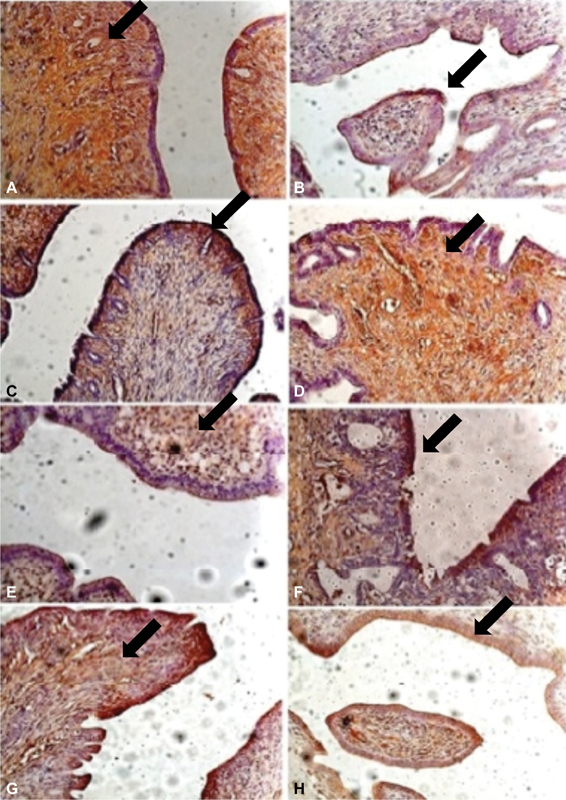
Objective: To characterize the patterns of cell differentiation, proliferation, and tissue invasion in eutopic and ectopic endometrium of rabbits with induced endometriotic lesions via a well- known experimental model, 4 and 8 weeks after the endometrial implantation procedure.
Methods: Twenty-nine female New Zealand rabbits underwent laparotomy for endometriosis induction through the resection of one uterine horn, isolation of the endometrium, and fixation of tissue segment to the pelvic peritoneum. Two groups of animals (one with 14 animals, and the other with15) were sacrificed 4 and 8 weeks after endometriosis induction. The lesion was excised along with the opposite uterine horn for endometrial gland and stroma determination. Immunohistochemical reactions were performed in eutopic and ectopic endometrial tissues for analysis of the following markers: metalloprotease (MMP-9) and tissue inhibitor of metalloprotease (TIMP-2), which are involved in the invasive capacity of the endometrial tissue; and metallothionein (MT) and p63, which are involved in cell differentiation and proliferation.
Results: The intensity of the immunostaining for MMP9, TIMP-2, MT, and p63 was higher in ectopic endometria than in eutopic endometria. However, when the ectopic lesions were compared at 4 and 8 weeks, no significant difference was observed, with the exception of the marker p63, which was more evident after 8 weeks of evolution of the ectopic endometrial tissue.
Conclusion: Ectopic endometrial lesions seem to express greater power for cell differentiation and tissue invasion, compared with eutopic endometria, demonstrating a potentially invasive, progressive, and heterogeneous presentation of endometriosis.
Objetivo: Caracterizar o padrão de diferenciação celular, proliferação e invasão tecidual em endométrio eutópico e ectópico de coelhas com lesões de endometriose induzidas por um modelo experimental 4 e 8 semanas após o procedimento de implantação endometrial. MéTODOS: Vinte e nove coelhas fêmeas Nova Zelândia foram submetidas a laparotomia para indução de endometriose através da ressecção de um dos cornos uterinos, isolamento do endométrio e fixação do tecido no peritônio pélvico. Dois grupos de animais (14 animais em um grupo e 15 animais no outro) foram sacrificados 4 e 8 semanas após a indução da endometriose. A lesão foi excisada junto com o corno uterino contralateral para determinação da presença de glândulas e de estroma endometrial. Reações de imunohistoquímica foram realizadas no tecido endometrial eutópico e ectópico para análise dos seguintes marcadores: metaloprotease (MMP9) e inibidor tecidual da metaloprotease 2 (TIMP-2), os quais estão envolvidos na capacidade de invasão do tecido endometrial; e metalotioneina (MT) e p63, os quais estão envolvidos na diferenciação e proliferação celular.
Resultados: A intensidade da imunomarcação para MMP9, TIMP-2, MT e p63 foi mais alta nos endométrios ectópicos do que nos endométrios eutópicos. Contudo, quando as lesões foram comparadas entre 4 e 8 semanas, nenhuma diferença foi observada, com exceção do marcador p63, o qual foi mais evidente depois de 8 semanas de evolução do tecido endometrial ectópico. CONCLUSãO: Lesões endometriais ectópicas parecem expressar maior poder de diferenciação celular e de invasão tecidual comparadas com endométrios eutópicos, demonstrando o potencial de invasão, de progressão e de apresentação heterogênea da endometriose.
Thieme Revinter Publicações Ltda Rio de Janeiro, Brazil.
Conflict of interest statement
The authors have no conflicts of interest to declare.
Figures
Similar articles
-
Increased cell proliferation in experimentally induced endometriosis in rabbits.Fertil Steril. 2010 Mar 15;93(5):1637-42. doi: 10.1016/j.fertnstert.2009.01.126. Epub 2009 Mar 21. Fertil Steril. 2010. PMID: 19304285
-
Matrix metalloproteinase-9 and tissue inhibitor of metalloproteinase-3 mRNA expression in ectopic and eutopic endometrium in women with endometriosis: a rationale for endometriotic invasiveness.Fertil Steril. 2001 Jan;75(1):152-9. doi: 10.1016/s0015-0282(00)01670-8. Fertil Steril. 2001. PMID: 11163831
-
[Adhesive and invasive effects and mechanism of neiyi recipe on the endometriosis].Zhongguo Zhong Xi Yi Jie He Za Zhi. 2011 Aug;31(8):1113-7. Zhongguo Zhong Xi Yi Jie He Za Zhi. 2011. PMID: 21910347 Chinese.
-
The role of endoplasmic reticulum stress in endometriosis.Cell Stress Chaperones. 2023 Mar;28(2):145-150. doi: 10.1007/s12192-023-01323-2. Epub 2023 Jan 25. Cell Stress Chaperones. 2023. PMID: 36696012 Free PMC article. Review.
-
Research Progress of Caspase in Endometriosis.Reprod Sci. 2024 Jun;31(6):1496-1507. doi: 10.1007/s43032-023-01425-3. Epub 2024 Feb 12. Reprod Sci. 2024. PMID: 38347381 Review.
Cited by
-
The Known, the Unknown and the Future of the Pathophysiology of Endometriosis.Int J Mol Sci. 2024 May 27;25(11):5815. doi: 10.3390/ijms25115815. Int J Mol Sci. 2024. PMID: 38892003 Free PMC article. Review.
-
p63 expression in granulosa-luteinized cells of infertile patients with peritoneal endometriosis submitted to in vitro fertilization.JBRA Assist Reprod. 2022 Aug 4;26(3):422-425. doi: 10.5935/1518-0557.20210090. JBRA Assist Reprod. 2022. PMID: 34713686 Free PMC article.
References
-
- Augoulea A, Alexandrou A, Creatsa M, Vrachnis N, Lambrinoudaki I.Pathogenesis of endometriosis: the role of genetics, inflammation and oxidative stress Arch Gynecol Obstet 20122860199–103. Doi: 10.1007/s00404-012-2357-8 - PubMed
-
- Chung H W, Wen Y, Chun S H, Nezhat C, Woo B H, Lake Polan M.Matrix metalloproteinase-9 and tissue inhibitor of metalloproteinase-3 mRNA expression in ectopic and eutopic endometrium in women with endometriosis: a rationale for endometriotic invasiveness Fertil Steril 20017501152–159. Doi: 10.1016/S0015-0282(00)01670-8 - PubMed
-
- Chung H W, Lee J Y, Moon H Set al.Matrix metalloproteinase-2, membranous type 1 matrix metalloproteinase, and tissue inhibitor of metalloproteinase-2 expression in ectopic and eutopic endometrium Fertil Steril 20027804787–795. Doi: 10.1016/S0015-0282(02)03322-8 - PubMed
-
- Uzan C, Cortez A, Dufournet C, Fauvet R, Siffroi J P, Daraï E.Eutopic endometrium and peritoneal, ovarian and bowel endometriotic tissues express a different profile of matrix metalloproteinases-2, -3 and -11, and of tissue inhibitor metalloproteinases-1 and -2 Virchows Arch 200444506603–609. Doi: 10.1007/s00428-004-1117-y - PubMed
MeSH terms
Substances
LinkOut - more resources
Full Text Sources
Medical
Research Materials
Miscellaneous