Review
. 2018 Nov 26;19(12):3752.
doi: 10.3390/ijms19123752.

Regenerative Models for the Integration and Regeneration of Head Skeletal Tissues

Affiliations
Review

Regenerative Models for the Integration and Regeneration of Head Skeletal Tissues

Warren A Vieira et al. Int J Mol Sci. .

Abstract

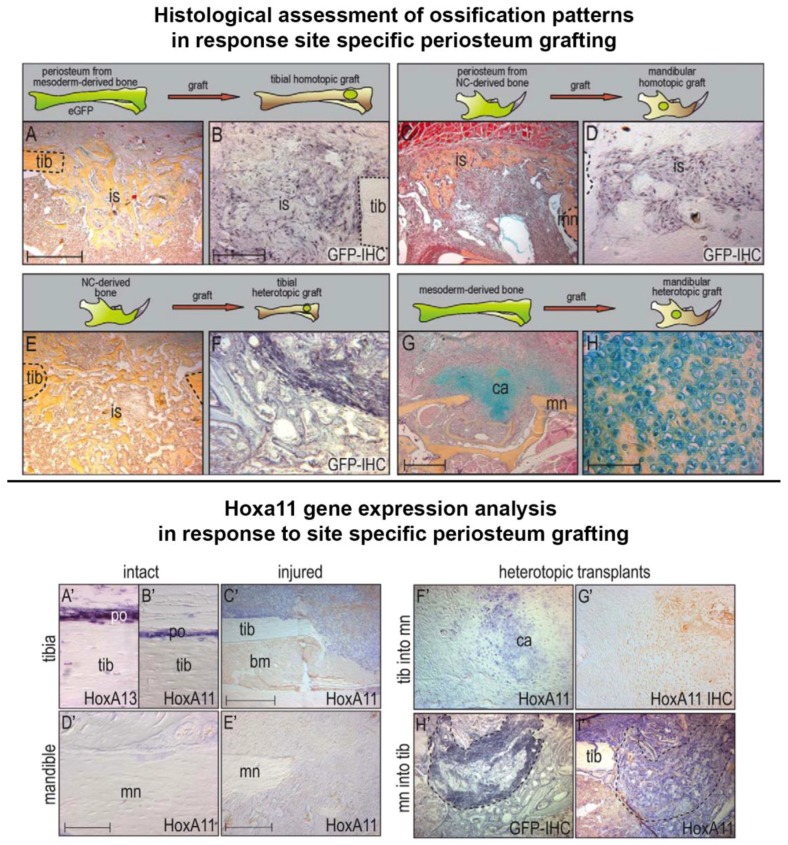
Disease of, or trauma to, the human jaw account for thousands of reconstructive surgeries performed every year. One of the most popular and successful treatment options in this context involves the transplantation of bone tissue from a different anatomical region into the affected jaw. Although, this method has been largely successful, the integration of the new bone into the existing bone is often imperfect, and the integration of the host soft tissues with the transplanted bone can be inconsistent, resulting in impaired function. Unlike humans, several vertebrate species, including fish and amphibians, demonstrate remarkable regenerative capabilities in response to jaw injury. Therefore, with the objective of identifying biological targets to promote and engineer improved outcomes in the context of jaw reconstructive surgery, we explore, compare and contrast the natural mechanisms of endogenous jaw and limb repair and regeneration in regenerative model organisms. We focus on the role of different cell types as they contribute to the regenerating structure; how mature cells acquire plasticity in vivo; the role of positional information in pattern formation and tissue integration, and limitations to endogenous regenerative and repair mechanisms.

Keywords: axolotl; jaw regeneration; limb regeneration; positional information; regenerative medicine; transplantation; zebrafish.


Conflict of interest statement

The authors declare no conflict of interest.

Figures

Figure 1
Figure 1
Whole mount preparations demonstrate the jaw-regenerative capacities of Urodeles and Zebrafish. Stage 40 newt larvae (top panel) exhibit more robust regenerative capabilities relative to their adult counter parts (middle panel) as well as to zebrafish (lower panel). In all three cases a cartilage rod is initially generated, fusing the two damaged ends of the mandible; however, this cartilage is replaced completely by bone in the zebrafish and results in the loss of the mandibular symphysis (MS). In the larval newt the regenerated pattern is identical to the original jaw; while in the adult regenerate is inappropriately patterned in terms of bone and cartilage relative to the original. Blue—cartilage staining, red—bone staining. Arrow in upper panel highlights the absence of dentary bone 13 dpa. Arrows in the middle panel highlights the dentary bone growing towards the mid-line 6wpa, and the absence of ossification on the lingual side of the jaw at 12 wpa. Dpa—days post amputation, wpa—weeks post amputation. (Images modified with permission from Ghosh et al. 1994 and Wang et al. 2012).
Figure 2
Figure 2
Differences and similarities in blastema and structure formation during adult Urodele jaw and limb regeneration. (A) The stages of jaw regeneration. The intact amphibian jaw consists of Meckel’s cartilage, dentary and prearticular bone, and the median symphysis connecting both sides of the jaw. The hyoid and brachial skeletons have been removed for simplicity. Upon amputation, a wound epithelium forms over the remaining jaw stump tissue. Independent of nerve singling, blastema cells accumulate at the severed edge of the jaw, derived from a variety of mature stump tissues, and will proliferate, repattern, and differentiate into a structure similar to the original structure, but with notable differences. The medium symphysis is lost and the majority of regenerated skeleton is cartilaginous (connect with Meckel’s cartilage), with only the dentary bone being restored. (B) The stages of limb regeneration. Limb regeneration also requires blastema formation, however its induction is slightly different from that of jaw regeneration. After amputation, a wound epithelium (WE) forms over the severed edge of the limb stump. The WE becomes innervated by the regenerating neurons, and establishes a specialized wound epithelium known as the Apical Epithelial Cap (AEC). The AEC is required for the dedifferentiation of the limb stump cells into blastema cells. The blastema cells, also derived from a variety of mature tissue, proliferate, pattern, and redifferentiate into all of the missing limb structures. The resulting limb regenerate is identical to the original.
Figure 3
Figure 3
Plasticity of tissue polarity during jaw and limb regeneration is conserved. (A) Jaw regeneration normally occurs with a posterior to anterior polarity; (B) When a portion of the jaw is reversed along its anterior-posterior axis and amputated, rather than generating structures that are posterior to the amputation plane, the blastema cells reverse their polarity so that they only regenerate structures anterior to the amputation plane. Dental lamina migrates into the adult jaw regenerate in a unidirectional manner, from posterior to anterior; however, the dental lamina migrates into the regenerate in the appropriate direction, despite the grafted tissue being rotated relative to the host site and regenerate; (C) During normal limb regeneration, the regenerate retains the proximal/distal polarity of the original limb; (D) Limbs with reversed proximal/distal axes can be generated by attaching the autopod to the flank. When the limb is amputated, the part of the limb that remains attached to the flank has the reverse proximal/distal orientation. However, rather than regenerating limb structures proximal to the wound site, the blastema reverses its polarity such that it regenerates tissue distal to the amputation plane. Therefore, there is a conserved mechanism, which prevents regeneration in a proximal direction. Red dashed lines represent the plane of amputation. Blue arrows indicate the direction of tissue polarity. Purple and orange lines represent dental lamina. Green arrow represents tissue grafting. Black arrows represent amputation and regeneration time.
Figure 4
Figure 4
Hox gene re-expression is a characteristic of regeneration shared between species and anatomical locations. Jaw, limb and tail blastema tissue exhibit the re-expression of both developmentally related and unrelated Hox genes. Red line indicates plan of amputation; blue tissue indicates blastema tissue.
Figure 5
Figure 5
The capacity of bone derived mesenchymal cells to participate in bone restoration is dependent on both the donor and recipient site. Top panel: Robust tibia bone repair, whereby pentachrome staining indicates intramembranous ossification (A), is facilitated directly by the homographic translation, survival and differentiation of GFP-positive tibial periosteum at the site of injury (B, GFP immunohistochemistry). A similar response is documented in mandibular sites (C) transplanted with GFP-positive mandibular periosteum (D). Although intramembranous bone is generated in tibial sites (E) in response to engraftment, survival and differentiation of GFP positive mandibular derived periosteum cells (F); tibial periosteum transplanted into the mandible exhibits failed integration due to inappropriate endochondral ossification (G) and cartilage condensation (H, high magnification). ca, cartilage; is, injury site; mn, mandible; tib, tibia. Scale bar: 200 µm in A, C, E; 100 µm in B, D, F; 400 µm in G; 50 µm in H. Lower panel: Periosteum derived from the adult tibia expresses Hoxa13 (A’) and Hoxa11 (B’) under homeostatic conditions, and Hoxa11 in response to injury (C’). Hoxa11 expression is absent in mandibular periosteum under both normal and injury conditions (D’,E’ respectively). Tibia periosteum retains Hoxa11 expression when transplanted into the Hox-negative mandibular environment (F’,G’), while GFP-positive mandibular periosteum (H’, GFP expression encompassed by dashed lines) grafted into the tibia exhibits Hoxa11 expression (I, mandibular cells encompassed by dashed lines) bm, bone marrow; ca, cartilage; mn, mandible; po, periosteum; tib, tibia. Scale bar: 50 µm in A, B, D, F, G; 100 µm in E; 200 µm in C, H, I. (Images republished with permission from Leucht et al. 2008).

Similar articles

Cited by

References

    1. Cawood J.I., Howell R.A. A classification of the edentulous jaws. Int. J. Oral Maxillofac. Surg. 1988;17:232–236. doi: 10.1016/S0901-5027(88)80047-X. - DOI - PubMed
    1. McCarthy J.G. The role of distraction osteogenesis in the reconstruction of the mandible in unilateral craniofacial microsomia. Clin. Plast. Surg. 1994;21:625–631. - PubMed
    1. Shwyrkow M.B., Shamsudinov A.K. Methods of simultaneous treatment of the mandible defects and the adjacent soft tissues. Acta Chir. Plast. 1989;31:226–235. - PubMed
    1. Komisar A. Mandibular Reconstruction. Thieme; Stuttgart, Germany: 1997.
    1. Glenske K., Donkiewicz P., Kowitsch A., Milosevic-Oljaca N., Rider P., Rofall S., Franke J., Jung O., Smeets R., Schnettler R., Wenisch S S., Barbeck M. Applications of Metals for Bone Regeneration. Int. J. Mol. Sci. 2018;19:826. doi: 10.3390/ijms19030826. - DOI - PMC - PubMed

LinkOut - more resources