. 2018 Nov 26;10(12):1829.
doi: 10.3390/nu10121829.

Nobiletin Enhances Chemosensitivity to Adriamycin through Modulation of the Akt/GSK3β/β⁻Catenin/MYCN/MRP1 Signaling Pathway in A549 Human Non-Small-Cell Lung Cancer Cells

Affiliations

Nobiletin Enhances Chemosensitivity to Adriamycin through Modulation of the Akt/GSK3β/β⁻Catenin/MYCN/MRP1 Signaling Pathway in A549 Human Non-Small-Cell Lung Cancer Cells

Jeong Yong Moon et al. Nutrients. .

Abstract

Drug resistance is a major problem in the treatment of non-small-cell lung cancer (NSCLC). In this study, Kyoto Encyclopedia of Genes and Genomes (KEGG) pathway analysis was performed to identify the differentially expressed genes in Adriamycin (ADR)-resistant NSCLC A549/ADR cells compared with parental A549 cells. Among the tested phytochemicals, nobiletin (NBT) is able to overcome the ADR resistance of A549/ADR cells. NBT treatment decreased the expression of a neuroblastoma-derived MYC (MYCN) and multidrug resistance-associated protein 1 (MRP1) as well as downregulating Akt, GSK3β, and β-catenin. Consistent with these results, NBT treatment resulted in the accumulation of intracellular ADR. A combination index (CI) assay confirmed the synergistic effect of combined treatment with NBT and ADR in reducing the viability of A549/ADR cells (CI = 0.152). Combined treatment with NBT and ADR enhanced apoptosis in A549/ADR cells, as evidenced by increased caspase-3 activation, poly (ADP-ribose) polymerase (PARP) cleavage, and sub-G1 population compared to treatment with ADR alone. In vivo experiments using a mouse xenograft model revealed that combination therapy with NBT and ADR significantly reduced tumor volume by 84.15%. These data suggest that NBT can sensitize ADR-induced cytotoxicity against A549/ADR cells by inhibiting MRP1 expression, indicating that NBT could serve as an effective adjuvant agent for ADR-based chemotherapy in lung cancer.

Keywords: A549 human non-small-cell lung cancer cells; Adriamycin (ADR); multidrug resistance-associated protein 1 (MRP1); nobiletin (NBT).


Conflict of interest statement

The authors declare no conflict of interest.

Figures

Figure 1
Figure 1
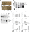
Comparison between A549 cells and ADR-resistant non-small cell lung cancer cells (A549/ADR). (A) MTT assay of A549 and A549/ADR cells treated with ADR (0.15–0.5 μM) for 48 h. (B) Morphological features of human lung A549 and A549/ADR cells treated with 0.5 µM ADR for 48 h. The cells were photographed at 200×. (C) A549 and A549/ADR cells were treated with ADR (0.5 μM) for 48 h and analyzed by flow cytometry after PI staining. The data shown are representative of three independent experiments indicating the quantification of sub-G1, G1, S, and G2/M populations. (D) Expression levels of apoptosis-related proteins were analyzed by Western blotting after 48 h of ADR treatment. β-actin was used as an internal control. The Western blotting intensities were quantified using ImageJ software. * p < 0.05. ADR: Adriamycin.
Figure 2
Figure 2
Analysis of differentially expressed genes (DEGs) in A549/ADR cells compared to A549 cells. (A) Heatmaps of gene expression data are shown for all samples. Representation of the log2-transformed values from 400 cellular genes identified as significantly altered in A549/ADR cells compared to A549 cells. (B) Top 10 pathways enriched by Gene Ontology (GO) analysis of DEGs in A549/ADR cells. The horizontal axis shows the percentage of annotated genes compared to the total gene number. The vertical axis shows the specific categories in each biological process, cellular component, and molecular function for GO as investigated by the Database for Annotation, Visualization and Integrated Discovery (DAVID) web tool and organized by p-values. The number shown on the right of each bar represents the number of genes, and the number in parentheses represents the p-value.
Figure 3
Figure 3
Top 10 altered pathways enriched in the Kyoto Encyclopedia of Genes and Genomes (KEGG) pathway analysis of A549/ADR cells. Pathways significantly enriched in (A) up-regulated genes, (B) down-regulated genes, and (C) total DEGs in A549/ADR cells vs. A549 cells analyzed in KEGG using DAVID. The horizontal axis shows the percentage of annotated genes compared to the total gene number. The vertical axis shows the pathways analyzed by KEGG analysis using DAVID. The pathways are organized by their p-values. The number shown on the right of each bar represents the number of genes, and the number in the parentheses represents the p-value. The red frame indicates the significant enrichment of the PI3K–Akt pathway from the KEGG pathway analysis.
Figure 4
Figure 4
Decrease in intracellular ADR accumulation in A549/ADR cells. (A) Representative confocal images of A549 and A549/ADR cells treated with 0.5 μM ADR (red) for 24 h. (B) The histogram shows the ADR accumulation in A549 cells and A549/ADR cells measured by flow cytometry. (C) Expression levels of ABC transporters were analyzed by Western blotting analysis in A549 and A549/ADR cells. The Western blot intensities were quantified using ImageJ software. * p < 0.05.
Figure 5
Figure 5
Nobiletin (NBT) enhances chemosensitivity of ADR by downregulating multidrug resistance-associated protein 1 (MRP1) expression through inhibiting Akt/GSK3β/β-catenin/ neuroblastoma-derived MYC (MYCN) in A549/ADR cells. (A) Cytotoxicity of ADR on A549/ADR cells treated with or without NBT as measured by MTT assay. (B) Combination index (CI) for various concentrations of NBT with ADR in A549/ADR cells. (C) Cytotoxicity of NBT in A549 and A549/ADR cells as measured by MTT assay. (D) MRP1 and Akt/GSK3β/β-catenin/MYCN signaling pathway expression levels in NBT-treated A549/ADR cells analyzed by Western blotting. (E) A549/ADR cells treated with NBT time dependently and analyzed by confocal immunofluorescence microscopy for Hoechst33342 (blue) and β-catenin (red) and their areas of overlap. Representative examples of three independent experiments are shown. Data are expressed as mean ± SD. * p < 0.05.
Figure 6
Figure 6
NBT enhances intracellular accumulation of ADR in A549/ADR cells. (A) Confocal microscopy images showing ADR accumulation in A549/ADR cells treated with 0.5 μM ADR alone (red) or ADR in combination with 50 μM NBT for 6, 12, and 24 h. (B) The histogram shows the relative fluorescence intensity of ADR uptake in A549/ADR cells treated with/without 0.5 μM ADR. (C) Intracellular 5-carboxyfluorescein (5-CF) retention as a measure of MRP1 activity in A549 and A549/ADR cells. (D) A549/ADR cells were treated with 5-carboxyfluorescein diacetate (5-CFDA) (10 μM) for 30 min with or without the addition of NBT (50 μM), MK571. * p < 0.05.
Figure 7
Figure 7
NBT enhances the apoptosis-initiating potential of ADR in A549/ADR cells. (A) Morphological features of A549/ADR cells after treatment with ADR (0.5 μM), NBT (50 μM), or both. (B) A549/ADR cells treated with ADR alone (0.5 μM), NBT alone (50 μM), and a combination of both were subjected to cell cycle analysis by flow cytometry using propidium iodide (PI) staining. (C) Expression levels of proteins were analyzed by Western blotting. β-actin was used as an internal control. (D) The intensities of the Western blotting bands were quantified using ImageJ software. * p < 0.05.
Figure 8
Figure 8
Antitumor effect of NBT in a nude mice xenograft tumor model. (A) Representative images of the tumor burden obtained from each group. (B) The tumor volume in each group was assessed by calipers and calculated as the length × width × width × 0.5. (C) Each bar represents the mean ± standard error of mean (SEM) of the tumor weight of four groups. (D) There were no significant changes in the body weights of the mice during the ADR and NBT treatment periods. * p < 0.05.

Similar articles

Cited by

References

    1. Torre L.A., Bray F., Siegel R.L., Ferlay J., Lortet-Tieulent J., Jemal A. Global cancer statistics, 2012. CA Cancer J. Clin. 2015;65:87–108. doi: 10.3322/caac.21262. - DOI - PubMed
    1. Ferlay J., Soerjomataram I., Ervik M., Dikshit R., Eser S., Mathers C., Rebelo M., Parkin D., Forman D., Bray F. Cancer Incidence and Mortality Worldwide: Iarc Cancer Base no. 11. International Agency for Research on Cancer; Paris, France: 2013.
    1. Cruz C.S.D., Tanoue L.T., Matthay R.A. Lung cancer: Epidemiology, etiology, and prevention. Clin. Chest Med. 2011;32:605–644. doi: 10.1016/j.ccm.2011.09.001. - DOI - PMC - PubMed
    1. Ramalingam S., Belani C. Systemic chemotherapy for advanced non-small cell lung cancer: Recent advances and future directions. Oncologist. 2008;13:5–13. doi: 10.1634/theoncologist.13-S1-5. - DOI - PubMed
    1. Le Chevalier T. Adjuvant chemotherapy for resectable non-small-cell lung cancer: Where is it going? Ann. Oncol. 2010;21:vii196–vii198. doi: 10.1093/annonc/mdq376. - DOI - PubMed

MeSH terms