Review
. 2018 Nov 26;19(12):3755.
doi: 10.3390/ijms19123755.

Functional Annotation of Bacterial Signal Transduction Systems: Progress and Challenges

Affiliations
Review

Functional Annotation of Bacterial Signal Transduction Systems: Progress and Challenges

David Martín-Mora et al. Int J Mol Sci. .

Abstract

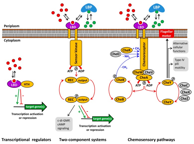
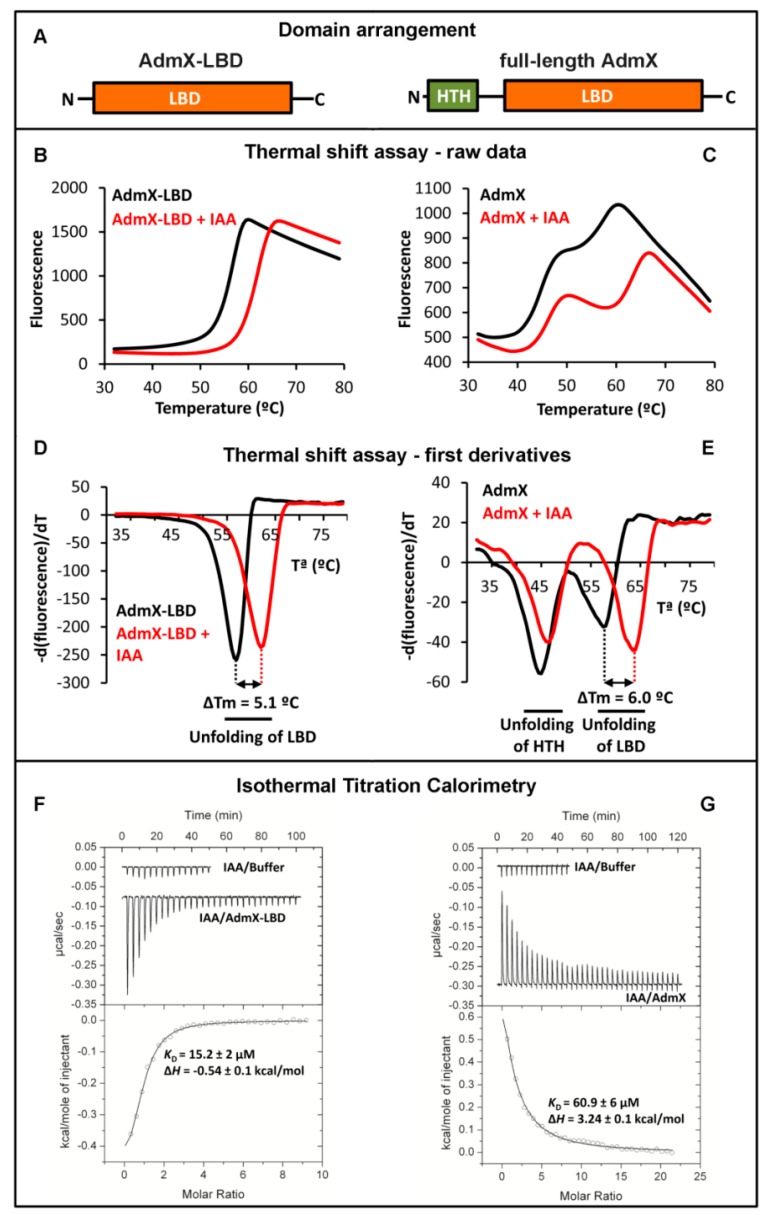
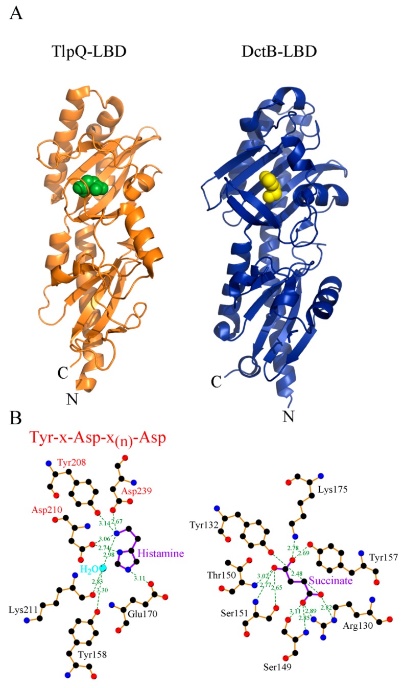
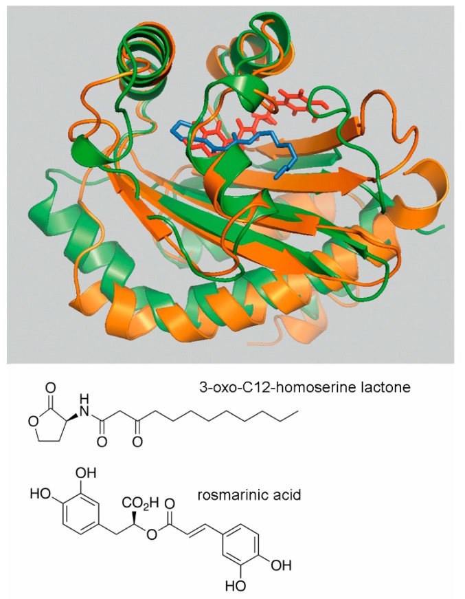
Bacteria possess a large number of signal transduction systems that sense and respond to different environmental cues. Most frequently these are transcriptional regulators, two-component systems and chemosensory pathways. A major bottleneck in the field of signal transduction is the lack of information on signal molecules that modulate the activity of the large majority of these systems. We review here the progress made in the functional annotation of sensor proteins using high-throughput ligand screening approaches of purified sensor proteins or individual ligand binding domains. In these assays, the alteration in protein thermal stability following ligand binding is monitored using Differential Scanning Fluorimetry. We illustrate on several examples how the identification of the sensor protein ligand has facilitated the elucidation of the molecular mechanism of the regulatory process. We will also discuss the use of virtual ligand screening approaches to identify sensor protein ligands. Both approaches have been successfully applied to functionally annotate a significant number of bacterial sensor proteins but can also be used to study proteins from other kingdoms. The major challenge consists in the study of sensor proteins that do not recognize signal molecules directly, but that are activated by signal molecule-loaded binding proteins.

Keywords: bacterial signal transduction systems; chemoreceptors; chemotaxis; sensor kinases; transcriptional regulators.


Conflict of interest statement

The authors declare no conflict of interest.

Figures

Figure 1
Figure 1
Major bacterial signal transduction systems. LBD: ligand binding domain; HTH: helix-turn-helix motif containing DNA binding domain; LBP: ligand binding protein; REC: receiver domain. The core proteins of chemosensory pathways, present in most pathways, are shaded in yellow, whereas auxiliary proteins, present only in some pathways, are shown in grey. red and green dots: signal molecules; red arrows represent phosphorylation/dephosphorylation events, blue arrows represent methylation/demethylation/deamidation of amino acids; black arrows indicate the pathway output, the green arrow and red T-bar indicate transcriptional activation or inhibition, respectively.
Figure 2
Figure 2
Use of the Thermal Shift Assay and Isothermal Titration Calorimetry to identify ligands that bind to AdmX. (A) Domain arrangement of AdmX and its ligand binding domain (LBD). (BE) Thermal shift experiments of AdmX and AdmX-LBD in the absence and presence of indole-3-acetic acid (IAA). (B,C) raw data; (D,E) first derivatives of raw data; (F,G) microcalorimetric titrations of buffer, AdmX and AdmX-LBD with IAA. Upper panel: titration raw data; lower panel: fit of dilution heat-corrected and concentration-normalized raw data with a model for the binding of a single ligand to a macromolecule. The derived thermodynamic binding parameters are indicated.
Figure 3
Figure 3
Three-dimensional structure and mode of ligand recognition at dCACHE domains. (A) Ribbon diagram of the LBD of the TlpQ chemoreceptor of P. aeruginosa (orange) in complex with histamine (green, pdb ID 6fu4) and LBD of the DctB sensor kinase of V. cholerae (blue) in complex with succinate (yellow, pdb ID 3by9). (B) Amino acids involved in ligand recognition. The sequence motif involved in amine recognition is shown in red above the histamine plot. Figure generated using Ligplot [76].
Figure 4
Figure 4
Use of virtual ligand screening to identify the plant compound rosmarinic acid as RhlR ligand. Superimposition of the ligand binding domains of LasR (green) in complex with 3-oxo-C12-homoserine lactone and the RhlR homology model (orange) onto which rosmarinic acid (RA) was docked. The LasR structure was obtained from the protein data bank (pdb ID 3IX3) and the RhlR model was generated by homology modelling as described in [83]. 3-oxo-C12-homoserine lactone and RA are shown in blue and red, respectively. The best binding position of RA with lowest glide score and glide energy is displayed. The structures of 3-oxo-C12-homoserine lactone and rosmarinic acid are shown in the lower part of the figure.
Figure 5
Figure 5
Sensor kinases that are activated by the binding of ligand loaded periplasmic binding proteins. Shown are structures of the LBDs of the sensor kinases LytS (pdb 5XSJ), TorS (pdb 3O1H) and LuxQ (pdb 2HJ9) (in red) in complex with their respective ligand-loaded binding proteins (in blue). The bacterial species and ligands bound (in yellow) are indicated.

Similar articles

Cited by

References

    1. Ulrich L.E., Koonin E.V., Zhulin I.B. One-component systems dominate signal transduction in prokaryotes. Trends Microbiol. 2005;13:52–56. doi: 10.1016/j.tim.2004.12.006. - DOI - PMC - PubMed
    1. Wuichet K., Zhulin I.B. Origins and diversification of a complex signal transduction system in prokaryotes. Sci. Signal. 2010;3:ra50. doi: 10.1126/scisignal.2000724. - DOI - PMC - PubMed
    1. Jacob-Dubuisson F., Mechaly A., Betton J.M., Antoine R. Structural insights into the signalling mechanisms of two-component systems. Nat. Rev. Microbiol. 2018;16:585–593. doi: 10.1038/s41579-018-0055-7. - DOI - PubMed
    1. Groisman E.A. Feedback Control of Two-Component Regulatory Systems. Annu. Rev. Microbiol. 2016;70:103–124. doi: 10.1146/annurev-micro-102215-095331. - DOI - PMC - PubMed
    1. Zschiedrich C.P., Keidel V., Szurmant H. Molecular Mechanisms of Two-Component Signal Transduction. J. Mol. Biol. 2016;428:3752–3775. doi: 10.1016/j.jmb.2016.08.003. - DOI - PMC - PubMed

LinkOut - more resources