. 2018 Nov 27;19(12):3763.
doi: 10.3390/ijms19123763.

Blood Outgrowth and Proliferation of Endothelial Colony Forming Cells are Related to Markers of Disease Severity in Patients with Pulmonary Arterial Hypertension

Affiliations

Blood Outgrowth and Proliferation of Endothelial Colony Forming Cells are Related to Markers of Disease Severity in Patients with Pulmonary Arterial Hypertension

Josien Smits et al. Int J Mol Sci. .

Abstract

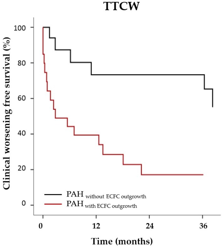
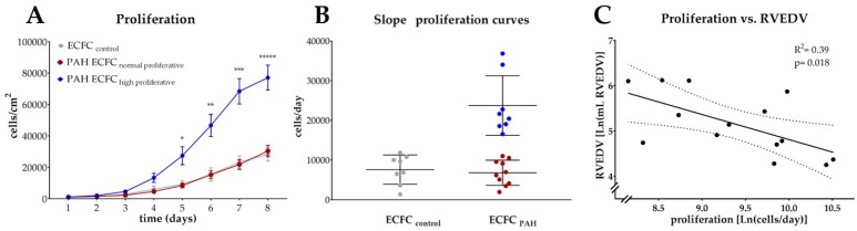
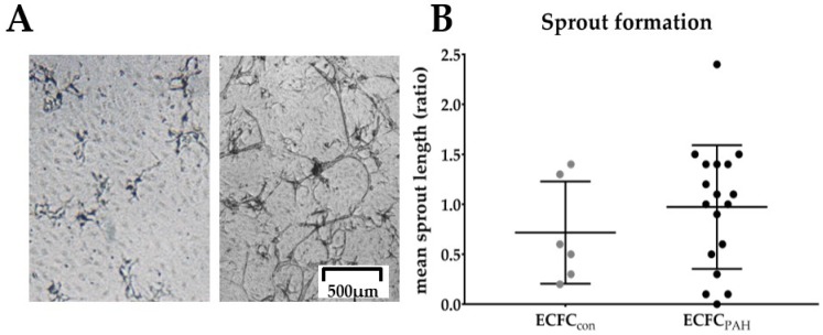
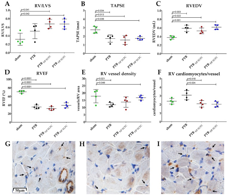
In pulmonary arterial hypertension (PAH), lung-angioproliferation leads to increased pulmonary vascular resistance, while simultaneous myocardial microvessel loss contributes to right ventricular (RV) failure. Endothelial colony forming cells (ECFC) are highly proliferative, angiogenic cells that may contribute to either pulmonary vascular obstruction or to RV microvascular adaptation. We hypothesize ECFC phenotypes (outgrowth, proliferation, tube formation) are related to markers of disease severity in a prospective cohort-study of 33 PAH and 30 healthy subjects. ECFC were transplanted in pulmonary trunk banded rats with RV failure. The presence of ECFC outgrowth in PAH patients was associated with low RV ejection fraction, low central venous saturation and a shorter time to clinical worsening (5.4 months (0.6⁻29.2) vs. 36.5 months (7.4⁻63.4), p = 0.032). Functionally, PAH ECFC had higher proliferative rates compared to control in vitro, although inter-patient variability was high. ECFC proliferation was inversely related to RV end diastolic volume (R² = 0.39, p = 0.018), but not pulmonary vascular resistance. Tube formation-ability was similar among donors. Normal and highly proliferative PAH ECFC were transplanted in pulmonary trunk banded rats. While no effect on hemodynamic measurements was observed, RV vascular density was restored. In conclusion, we found that ECFC outgrowth associates with high clinical severity in PAH, suggesting recruitment. Transplantation of highly proliferative ECFC restored myocardial vascular density in pulmonary trunk banded rats, while RV functional improvements were not observed.

Keywords: endothelial colony forming cell; endothelial progenitor cell; pulmonary hypertension; pulmonary vascular disease.


Conflict of interest statement

The authors declare no conflict of interest.

Figures

Figure 1
Figure 1
PAH patients with outgrowth of ECFC are more likely to experience early clinical worsening. Kaplan Meijer curves of PAH patients with outgrowth of ECFC (red, n = 21) without outgrowth of ECFC (grey, n = 12) over a period of 36 months (x-axis). Cox-regression corrected for age; p = 0.032.
Figure 2
Figure 2
PAH ECFC have higher proliferative rates compared to ECFC from healthy control subjects, while ECFC proliferation is related to RVEDV. (A) Quantification of cell density (y-axis, cells/cm2) of ECFC of healthy subjects (n = 8, grey) and PAH donors (n = 18, red and blue) over time (x-axis, days) indicates PAH ECFC proliferation is significantly higher compared to ECFC from healthy subjects, due to a subset of highly proliferative of PAH ECFC (n = 8, blue). Two way repeated measurements ANOVA, p < 0.0001. Mean with standard error of mean (SEM) is shown. (B) Quantification of the steepness of individual growth curves (y-axis, cells/day) indicates high proliferative PAH ECFC exceed the mean steepness of the growth curve of control ECFC by >2SD, where mean with SD is shown. (C) Individual steepness of the growth curve of PAH ECFC is correlated to RVEDV (y-axis, Ln (RVEDV, mL), linear regression, R2 = 0.39, p = 0.018, n = 14. 95% confidence interval (CI) is shown of Ln transformed data.
Figure 3
Figure 3
No difference in tube formation was observed among ECFC donors. (A) Representative phase contrast image of sprouts of PAH ECFC showing little or abundant tube formation. Scale bar indicates 500 μm. (B) Quantitative analysis of average tube length indicates no difference between control (grey, n = 6) or PAH ECFC (black, n = 18), normalized for mean tube length of control ECFC (y-axis, ratio). Mean with standard deviation (SD) is shown.
Figure 4
Figure 4
Transplantation of PAH ECFC did not induce reversal of the hemodynamic profile in PTB rats of chronic RV failure. (A) RV/LVS weight-ratio was comparable among sham rats (0.32 ± 0.14, green), PTB rats (0.51 ± 0.18, black), PTB rats transplanted with normal proliferative PAH ECFC (0.67 ± 0.11, red), or PTB rats transplanted with highly proliferative PAH ECFC (0.67 ± 0.11, blue). (B) TAPSE at evaluation shown of sham (2.7 ± 0.3 mm), PTB (1.7 ± 0.3 mm), PTB normal proliferative PAH ECFC (1.6 ± 0.5 mm), or PTB high proliferative PAH ECFC (1.7 ± 0.19 mm). (C) RVEDV of sham (0.38 ± 0.04 mL), PTB (0.59 ± 0.07 mL), PTB normal proliferative PAH ECFC (0.53 ± 0.07 mL) and PTB high proliferative PAH ECFC (0.62 ± 0.06 mL). (D) RVEF of sham (71 ± 3%), PTB (36 ± 5%), PTB normal proliferative PAH ECFC (32 ± 4%) and PTB high proliferative PAH ECFC (39 ± 6%). (E) Quantification of small vessel density/RV-area indicates significant reversal of the RV myocardial vessel density after transplantation with highly proliferative PAH ECFC (16.8 ± 1.6 vessels/RV-area), sham-animals (17.8 ± 4.6 vessels/RV-area), PTB (11.9 ± 1.5 vessels/RV-area), PTB normal proliferative PAH ECFC (13.7 ± 3.6 vessels/RV-area. (F) Cardiomyocytes per vessel ratio for sham (2.4 ± 0.4), PTB (3.1 ± 0.5), PTB normal proliferative PAH ECFC (2.0 ± 0.5) and high proliferative PAH ECFC (2.1 ± 0.6). Mean with SD is shown. Reported p-values are obtained from independent samples two way ANOVA with Bonferroni correction. (GI) Representative phase contrast images of RV-muscle with brown CD31 staining for vessels (black arrow is pointed at a vessel) and nuclei (DAPI) of sham rats (G), PTB banded rats (H) and PTB banded rats transplanted with highly proliferative PAH ECFC (I). Scale bar indicates 50 μm.

Similar articles

Cited by

References

    1. Voelkel N.F., Gomez-Arroyo J.G., Abbate A., Bogaard H.J., Nicolls M.R. Pathobiology of pulmonary arterial hypertension and right ventricular failure. Eur. Respir J. 2012;40:1555–1565. doi: 10.1183/09031936.00046612. - DOI - PMC - PubMed
    1. Guignabert C., Dorfmuller P. Pathology and pathobiology of pulmonary hypertension. Semin. Respir. Crit. Care Med. 2013;34:551–559. doi: 10.1055/s-0033-1356496. - DOI - PubMed
    1. Vonk-Noordegraaf A., Haddad F.O., Chin K.M., Forfia P.R., Kawut S.M., Lumens J., Naeije R., Newman J., Oudiz R.J., Provencher S., et al. Right Heart Adaptation to Pulmonary Arterial-Hypertension: Physiology and Pathobiology. J. Am. Coll. Cardiol. 2013;62:D22–D33. doi: 10.1016/j.jacc.2013.10.027. - DOI - PubMed
    1. Gomez-Arroyo J., Sandoval J., Simon M.A., Dominguez-Cano E., Voelkel N.F., Bogaard H.J. Treatment for pulmonary arterial hypertension-associated right ventricular dysfunction. Ann. Am. Thorac. Soc. 2014;11:1101–1115. doi: 10.1513/AnnalsATS.201312-425FR. - DOI - PubMed
    1. Rose J.A., Erzurum S., Asosingh K. Biology and flow cytometry of proangiogenic hematopoietic progenitors cells. Cytometry A. 2015;87:5–19. doi: 10.1002/cyto.a.22596. - DOI - PMC - PubMed

MeSH terms

Grants and funding