. 2018 Nov 27;7(12):489.
doi: 10.3390/jcm7120489.

Pterostilbene Attenuates Hexavalent Chromium-Induced Allergic Contact Dermatitis by Preventing Cell Apoptosis and Inhibiting IL-1β-Related NLRP3 Inflammasome Activation

Affiliations

Pterostilbene Attenuates Hexavalent Chromium-Induced Allergic Contact Dermatitis by Preventing Cell Apoptosis and Inhibiting IL-1β-Related NLRP3 Inflammasome Activation

Bour-Jr Wang et al. J Clin Med. .

Abstract

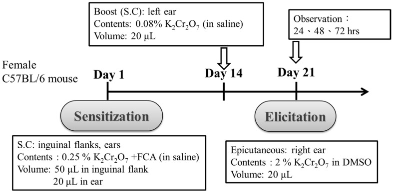
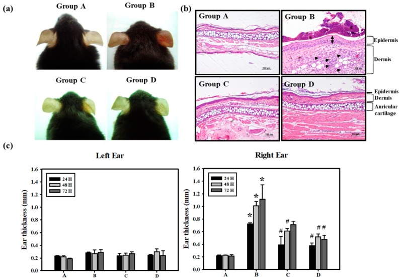
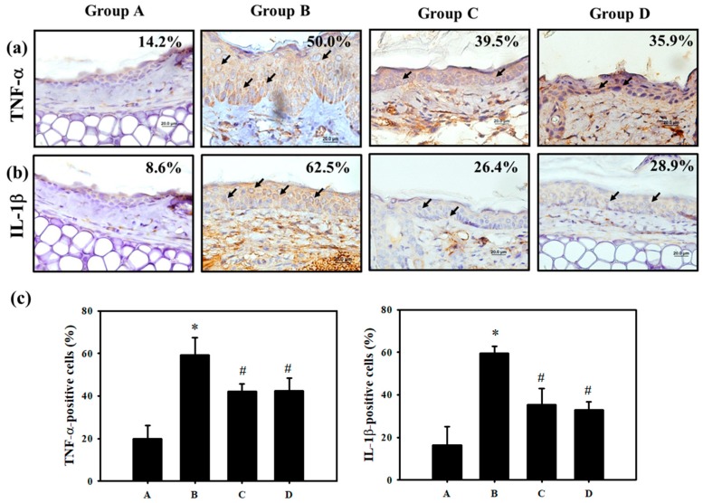
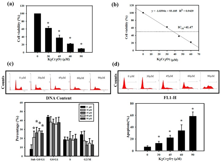
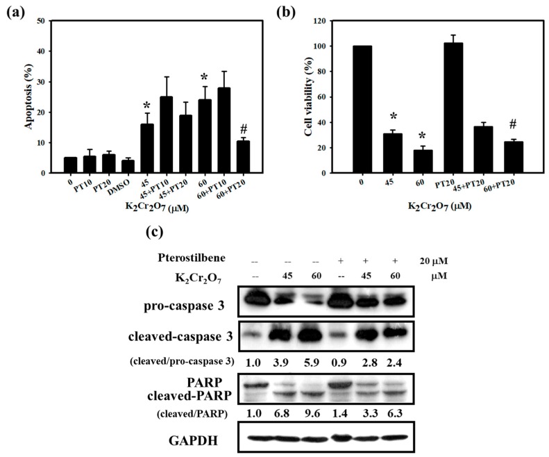
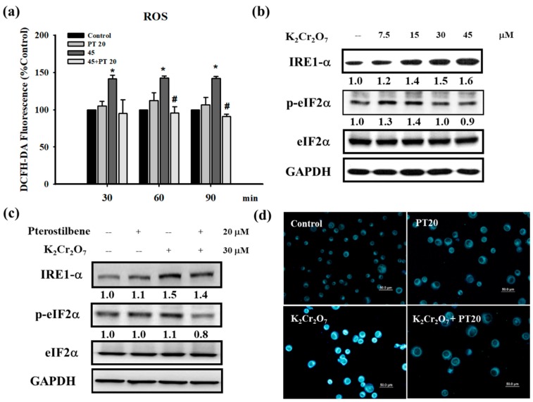
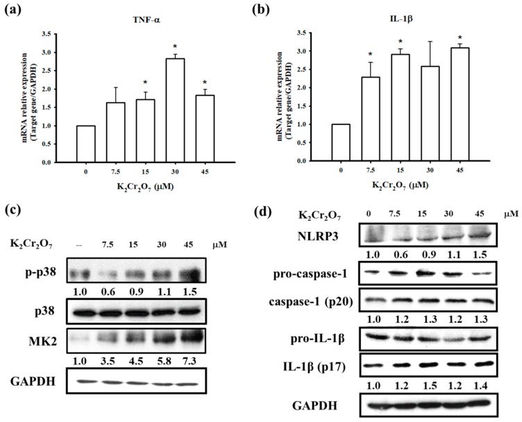
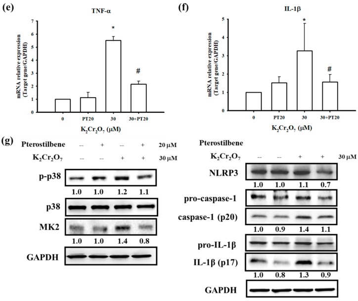
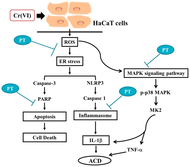
Hexavalent chromium (Cr(VI)) is widely used in many industries but can induce contact dermatitis especially in cement industries. Many cement workers suffer from Cr(VI)-induced allergic contact dermatitis (ACD), and prevention and therapeutic strategies are still lacking. Pterostilbene (PT) is a natural compound predominantly found in blueberries. Studies indicate the potential use of PT as an effective anti-oxidative and anti-inflammatory agent. Herein, we investigated the possible mechanisms involved and whether chromium-induced ACD could be effectively inhibited by treating PT. In our in vivo study, epidermal Cr(VI) administration causes cutaneous inflammation in mice ear skin, and the pro-inflammatory cytokines, TNF-α and IL-1β, were found in the epidermis, presenting the level of increase after Cr(VI) treatment. Meanwhile, the results of our in vitro experiment showed that apoptosis and endoplasmic reticulum (ER) stress were induced after treatment with different concentrations of Cr(VI) in HaCaT cells (human keratinocyte). Cr(VI) also induced TNF-α and IL-1β mRNA expressions, through the activation of the p38 mitogen-activated protein kinase (MAPK)/MAPK-activated protein kinase 2 (MK2) pathway. Notably, the severity of the skin reactions in the epicutaneous elicitation test significantly diminished when the mouse was treated with PT. Likewise, PT intervention also ameliorated the inflammation and apoptosis of HaCaT cells in vitro. Furthermore, our current findings demonstrated that the NLRP3 inflammasome could be involved in the Cr(VI)-mediated inflammation and apoptosis of ACD. Thus, interrupting this mechanism with proper nontoxic agents, such as PT, could be a new option to improve occupational chromium toxicity and hypersensitivity.

Keywords: NLRP3 inflammasome; allergic contact dermatitis; endoplasmic reticulum stress; hexavalent chromium; inflammatory cytokines; pterostilbene.


Conflict of interest statement

The authors declare no conflict of interest.

Figures

Figure 1
Figure 1
Scheme of intradermal sensitization and epicutaneous elicitation in an allergic contact dermatitis mouse model. S.C.: subcutaneous injection.
Figure 2
Figure 2
Protective effects of PT on Cr(VI)-induced ACD mice. (a) Swelling and erythema were documented by photography at 48 h. (b) H&E histology of mice ear skin after the elicitation test with Cr(VI) showed ear swelling and inflammatory cell infiltration at 72 h. In the dermis, the increased number of inflammatory cells, neutrophils, was observed (arrowhead) in K2Cr2O7 treatment (Group B). In the epidermis, Group B showed the stratum corneum thickening (bi-arrowhead), but NAC (Group C) or PT (Group D) treatment significantly suppressed Cr(VI)-induced increases in ear thickness. Also, no differences in the auricular cartilage were observed (scale bar representing 100 μM). (c) ACD reactions were quantified by measuring the ear thickness at 24, 48 and 72 h after the elicitation test (n = 5). The time course measurement of Left (vehicle) and Right (epicutaneous) ear thickness. (A: Control; B: K2Cr2O7; C: NAC; D: PT) (* p <0.05 versus control group; # p <0.05 versus K2Cr2O7 group).
Figure 3
Figure 3
PT protects against pro-inflammatory cytokines in mice with ACD induced by Cr(VI). Skin biopsy from the Control, K2Cr2O7, NAC and PT groups shows immunohistochemical staining for analysis of (a) TNF-α. (b) IL-1β- positive cells (brown) in the epidermis (scale bar representing 20 μM). (c) The quantification of TNF-α and IL-1β-positive cells was determined using HistoQuest software (Version 4.0.4.0158, TissueGnostics). (n = 3, * p <0.05 versus control group; # p <0.05 versus K2Cr2O7 group).
Figure 4
Figure 4
Measurement of cell viability, cell cycle distribution and apoptosis of Cr(VI) in HaCaT cells. The cells were treated with 0, 30, 45, 60 or 90 μM Cr(VI) for 24 h. (a) Cell viability was measured using the MTT assay. Cr(VI) decreased the viability of HaCaT cells in a concentration-dependent manner. (b) Determination of Cr(VI) IC50 values by linear regression. (c) Cells were collected and incubated with 50 μg/mL of PI for 30 min and subjected to flow cytometry analysis to examine the cell distribution at each phase of the cell cycle. The percentage of cells in the sub G0/G1 phase were significantly increased when cells were exposed to Cr(VI). (d) Early apoptosis detection was measured by an Annexin-V apoptosis detection kit. Flow cytometry analysis indicating the rate of apoptosis in HaCaT cells treated with Cr(VI). Quantification of early apoptosis in HaCaT cells exposed to Cr(VI) in a concentration-dependent manner. All above data are presented as the mean ± standard deviation (SD) from three independent experiments. * p <0.05 versus untreated cells.
Figure 5
Figure 5
Pretreatment with PT rescued Cr(VI)-induced cytotoxicity. (a) HaCaT cells were pretreated with 10 or 20 μM PT for 1 h. Early apoptosis, detected using an Annexin V apoptosis detection kit, showed that PT (20 μM) inhibited Cr(VI)-induced apoptosis at different concentrations of Cr(VI) (45 and 60 μM) exposure for 24 h. (b) Cell viability was measured using MTT assay. HaCaT cells exposed to Cr(VI) for 24 h after pretreatment with 20 μM PT. PT slightly increased the cell viability of Cr(VI) (60 μM). Data are presented as the mean ± SD from three independent experiments. * p <0.05 versus control; # p <0.05 versus the Cr(VI)-treated group. (c) The expression of apoptosis proteins, pro-caspase 3, cleaved-caspase 3, PARP and GAPDH were monitored after 24 h of Cr(VI) or Cr(VI) plus PT exposure in HaCaT cells. Western blots are representative of n = 3 experiments.
Figure 6
Figure 6
Cr(VI) treatment induced ROS and ER stress in HaCaT cells, which were inhibited by PT. (a) HaCaT cells were treated with 45 μM of Cr(VI) for various time periods (30, 60, and 90 min). The amount of ROS produced in cells was determined using the fluorescence dye (DCFH-DA) and analyzed by flow cytometry. Pretreatment with PT (20 μM) could suppress ROS generation. Fluorescence units reflected the subtraction of untreated cell values and were presented as the mean ± SD from three independent experiments. * p <0.05 versus control; # p <0.05 versus the Cr(VI)-treated group. (b) Cell lysates extracted from HaCaT cells treated with several concentrations (7.5, 15, 30, and 45 μM) of Cr(VI) for 24 h were submitted to Western blot analysis to detect the protein expression of ER stress. Cr(VI) upregulated IRE1α and p-eIF2α protein expressions but p-eIF2α decreased at 45 μM of Cr(VI). (c) HaCaT cells were treated with 30 μM of Cr(VI) for 24 h. Pretreatment with PT (20 μM) inhibited ER stress-related protein expression. Western blots are representative of n = 3 experiments. (d) HaCaT cells were treated with Cr(VI) (30 μM) for 24 h. The strength of the ER tracker was enhanced by Cr(VI) and alleviated by PT pretreatment. The scale bars are 50 μM.
Figure 7
Figure 7
PT attenuates the Cr(VI)-induced pro-inflammatory cytokines and NLRP3 inflammasome. (a,b) The mRNA expressions of TNF-α and IL-1β were measured by real-time PCR. HaCaT cells were treated with several concentrations (7.5, 15, 30, and 45 μM) of Cr(VI) for 12 h. Data presented as the mean ± SD from three independent experiments. * p <0.05 versus controls. (c,d) The expression of the p38 MAPK/MK2 signaling pathway and NLRP3 inflammasome-associated proteins were measured by Western blot analysis following treatment with several concentrations of Cr(VI) for 24 h. (e,f) The cells were pretreated with PT (20 μM) for 1 h and then treated with Cr(VI) (30 μM) for an additional 12 h. Real-time PCR showed that the mRNA levels of pro-inflammatory cytokines, TNF-α and IL-1β decreased after treatment with PT. (g) Western blot analysis demonstrated that the activation of the p38 MAPK/MK2 signaling pathway and the NLRP3 inflammasome was suppressed by PT. Western blots are representative of n = 3 experiments.
Figure 7
Figure 7
PT attenuates the Cr(VI)-induced pro-inflammatory cytokines and NLRP3 inflammasome. (a,b) The mRNA expressions of TNF-α and IL-1β were measured by real-time PCR. HaCaT cells were treated with several concentrations (7.5, 15, 30, and 45 μM) of Cr(VI) for 12 h. Data presented as the mean ± SD from three independent experiments. * p <0.05 versus controls. (c,d) The expression of the p38 MAPK/MK2 signaling pathway and NLRP3 inflammasome-associated proteins were measured by Western blot analysis following treatment with several concentrations of Cr(VI) for 24 h. (e,f) The cells were pretreated with PT (20 μM) for 1 h and then treated with Cr(VI) (30 μM) for an additional 12 h. Real-time PCR showed that the mRNA levels of pro-inflammatory cytokines, TNF-α and IL-1β decreased after treatment with PT. (g) Western blot analysis demonstrated that the activation of the p38 MAPK/MK2 signaling pathway and the NLRP3 inflammasome was suppressed by PT. Western blots are representative of n = 3 experiments.
Figure 8
Figure 8
Possible molecular targets of the preventive effects of PT in Cr(VI)-induced ACD. Cr(VI) activated NLRP3 inflammasome activity and the p38 MAPK/MK2 signaling pathway leading to pro-inflammatory gene expression and resulting in keratinocyte cell death by apoptosis. The administration of PT inhibited cell death, the NLRP3 inflammasome and pro-inflammatory cytokines, which were regulated by ROS generation and ER stress and were related to Cr(VI)-induced ACD.

Similar articles

Cited by

References

    1. Shelnutt S.R., Goad P., Belsito D.V. Dermatological toxicity of hexavalent chromium. Crit. Rev. Toxicol. 2007;37:375–387. doi: 10.1080/10408440701266582. - DOI - PubMed
    1. Goh C.L., Gan S.L., Ngui S.J. Occupational dermatitis in a prefabrication construction factory. Contact Dermat. 1986;15:235–240. doi: 10.1111/j.1600-0536.1986.tb01343.x. - DOI - PubMed
    1. Guo Y.L., Wang B.J., Yeh K.C., Wang J.C., Kao H.H., Wang M.T., Shih H.C., Chen C.J. Dermatoses in cement workers in southern Taiwan. Contact Dermat. 1999;40:1–7. doi: 10.1111/j.1600-0536.1999.tb05968.x. - DOI - PubMed
    1. Toncic R.J., Lipozencic J., Martinac I., Greguric S. Immunology of allergic contact dermatitis. Acta Dermatovenerol. Croat. 2011;19:51–68. - PubMed
    1. Fuchs J., Zollner T.M., Kaufmann R., Podda M. Redox-modulated pathways in inflammatory skin diseases. Free Radic. Biol. Med. 2001;30:337–353. doi: 10.1016/S0891-5849(00)00482-2. - DOI - PubMed

LinkOut - more resources