

Age-Dependent Differential Expression of Apoptotic Markers in Rat Oral Mucosa
- PMID: 30486627
- PMCID: PMC6318412
- DOI: 10.31557/APJCP.2018.19.11.3245
Age-Dependent Differential Expression of Apoptotic Markers in Rat Oral Mucosa
Abstract
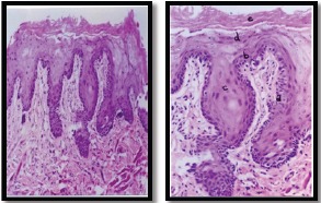
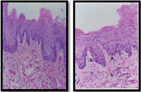
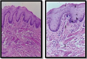
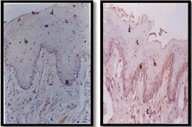
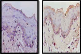
Objective: This study tests the hypothesis that gingival tissue produces age-dependent activation of apoptotic markers. Methods: To address the hypothesis, a prospective experimental study was conducted on 20 adult male albino rats, which were divided into two groups. Group 1 comprised rats aged six months (weighing 150–200 g), and group 2 included old rats aged one year (weighing 250–300 g). Gingival tissue and buccal mucosa biopsy samples were obtained from both groups. Histological and immunohistochemical (Bax apoptotic protein marker) sections were analyzed for both groups. Results: Our data showed a significant decrease in the proliferative activity of oral mucosa (gingiva and buccal mucosa) in old rats and an increase in the immunoreactivity of Bax apoptotic proteins related to increased susceptibility of cells to apoptosis. The mucosal structures (epithelium and lamina propria) were significantly different between the two groups. Furthermore, immunoreactivity for Bax was different between young and old rats. Conclusions: Aging is associated with changes that lead to progressive, irreversible deterioration of the functional capacities of several tissues and organs. Our study demonstrated the effect of age on the histological and apoptotic behavior of oral mucosa (gingiva and buccal mucosa) cells.
Keywords: Apoptosis; Bax; Bcl-2; gingival tissue.
Creative Commons Attribution License
Figures

Similar articles
-
Effects of aging on apoptosis gene expression in oral mucosal tissues.Apoptosis. 2013 Mar;18(3):249-59. doi: 10.1007/s10495-013-0806-x. Apoptosis. 2013. PMID: 23334583 Free PMC article.
-
Age-dependent differential expression of apoptosis markers in the gingival tissue.Arch Oral Biol. 2009 Apr;54(4):329-36. doi: 10.1016/j.archoralbio.2009.01.008. Epub 2009 Feb 12. Arch Oral Biol. 2009. PMID: 19215909
-
Immunoexpression of vascular endothelial growth factor in gingival mucosa with papilloma and condyloma acuminata.Rom J Morphol Embryol. 2015;56(3):1077-83. Rom J Morphol Embryol. 2015. PMID: 26662142
-
Parthenolide attenuates 7,12-dimethylbenz[a]anthracene induced hamster buccal pouch carcinogenesis.Mol Cell Biochem. 2018 Mar;440(1-2):11-22. doi: 10.1007/s11010-017-3151-5. Epub 2017 Aug 12. Mol Cell Biochem. 2018. PMID: 28801714
-
The effect of age on proliferating cell nuclear antigen expression in oral gingival epithelium of healthy and inflamed human gingiva.J Periodontol. 2000 Oct;71(10):1567-74. doi: 10.1902/jop.2000.71.10.1567. J Periodontol. 2000. PMID: 11063389
Cited by
-
Melatonin and Sirtuins in Buccal Epithelium: Potential Biomarkers of Aging and Age-Related Pathologies.Int J Mol Sci. 2020 Oct 30;21(21):8134. doi: 10.3390/ijms21218134. Int J Mol Sci. 2020. PMID: 33143333 Free PMC article.
-
Impact of age-related changes in buccal epithelial cells on pediatric epigenetic biomarker research.Nat Commun. 2025 Jan 12;16(1):609. doi: 10.1038/s41467-025-55909-8. Nat Commun. 2025. PMID: 39800776 Free PMC article.
References
-
- Andreescu CF, Mihai LL, Răescu M, et al. Age influence on periodontal tissues:A histological study. Rom J Morphol Embryol. 2013;54:811–5. - PubMed
-
- Borner C. The Bcl-2 protein family:sensors and checkpoints for life-or-death decisions. Mol Immunol. 2003;39:615–47. - PubMed
-
- Bryant R, Nix D. Acute and chronic wounds - E-Book - Ruth Bryant, Denise Nix Google Books. 2016
MeSH terms
Substances
LinkOut - more resources
Full Text Sources
Research Materials