. 2018 Dec 4;7(23):e010404.
doi: 10.1161/JAHA.118.010404.

Caspase Cleavage of Gelsolin Is an Inductive Cue for Pathologic Cardiac Hypertrophy

Affiliations

Caspase Cleavage of Gelsolin Is an Inductive Cue for Pathologic Cardiac Hypertrophy

Charis Putinski et al. J Am Heart Assoc. .

Abstract

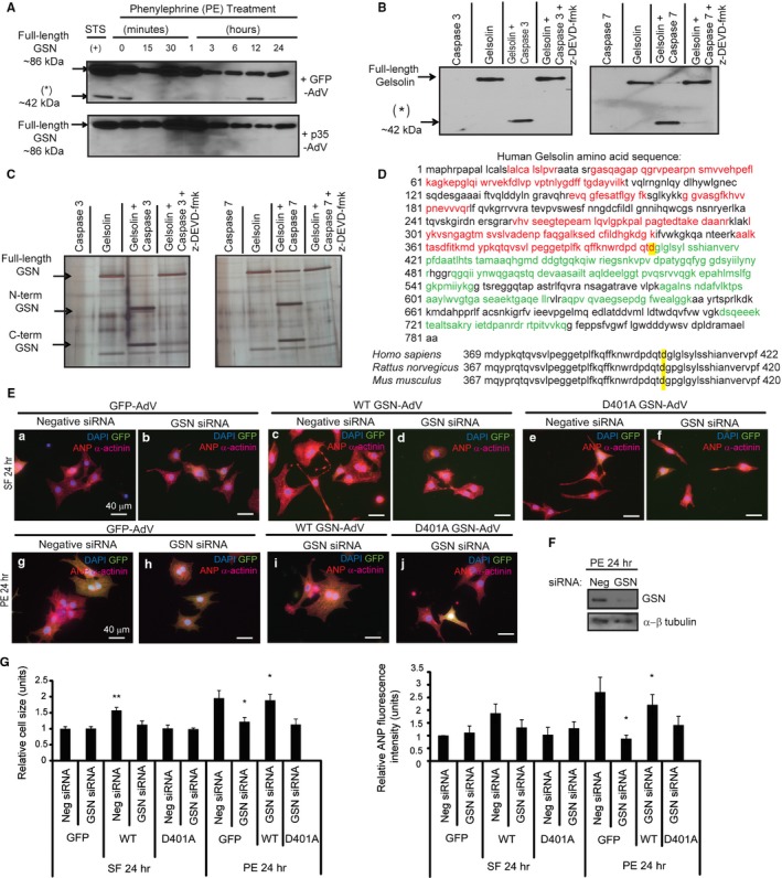
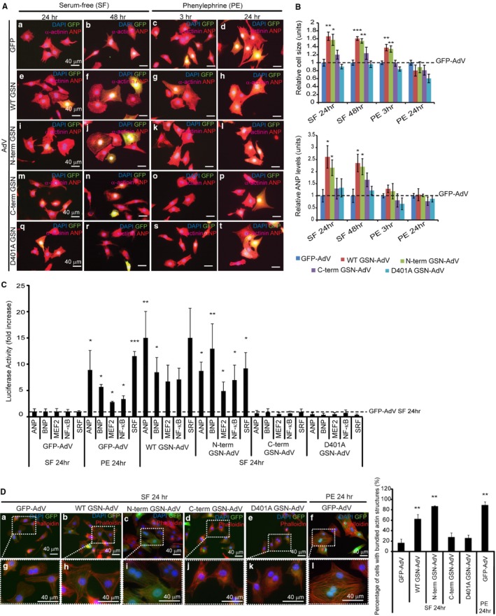
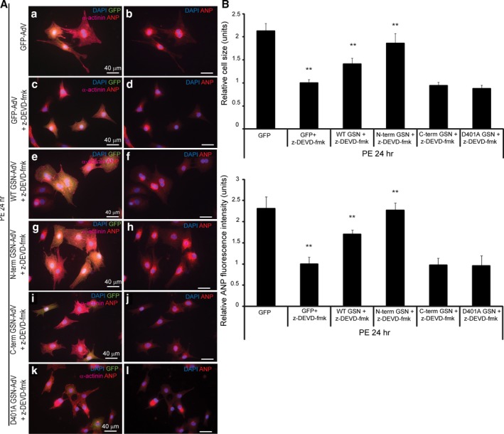
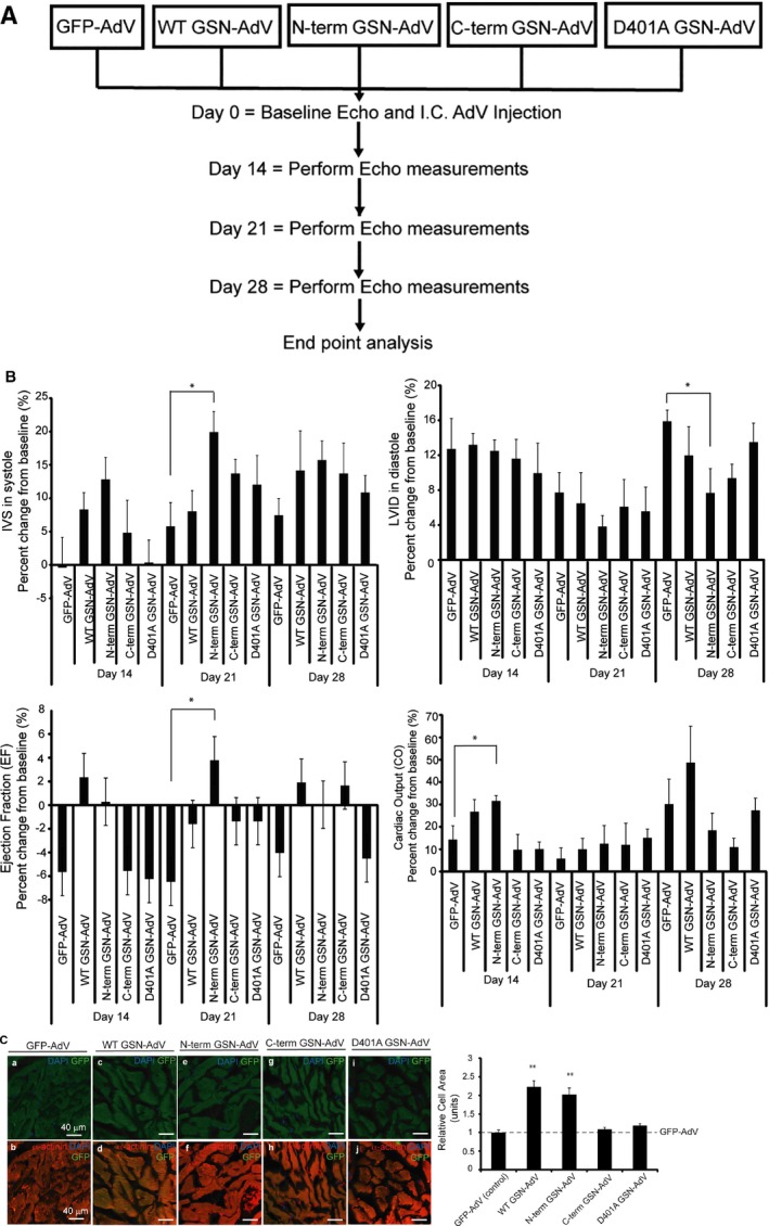
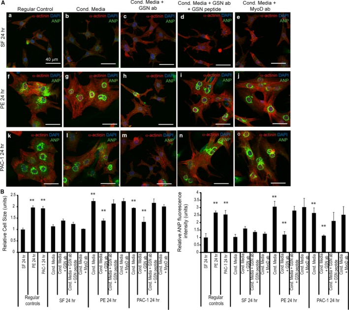
Background Cardiac hypertrophy is an adaptive remodeling event that may improve or diminish contractile performance of the heart. Physiologic and pathologic hypertrophy yield distinct outcomes, yet both are dependent on caspase-directed proteolysis. This suggests that each form of myocardial growth may derive from a specific caspase cleavage event(s). We examined whether caspase 3 cleavage of the actin capping/severing protein gelsolin is essential for the development of pathologic hypertrophy. Methods and Results Caspase targeting of gelsolin was established through protein analysis of hypertrophic cardiomyocytes and mass spectrometry mapping of cleavage sites. Pathologic agonists induced late-stage caspase-mediated cleavage of gelsolin. The requirement of caspase-mediated gelsolin cleavage for hypertrophy induction was evaluated in primary cardiomyocytes by cell size analysis, monitoring of prohypertrophy markers, and measurement of hypertrophy-related transcription activity. The in vivo impact of caspase-mediated cleavage was investigated by echo-guided intramyocardial injection of adenoviral-expressed gelsolin. Expression of the N-terminal gelsolin caspase cleavage fragment was necessary and sufficient to cause pathologic remodeling in isolated cardiomyocytes and the intact heart, whereas expression of a noncleavable form prevents cardiac remodeling. Alterations in myocardium structure and function were determined by echocardiography and end-stage cardiomyocyte cell size analysis. Gelsolin secretion was also monitored for its impact on naïve cells using competitive antibody trapping, demonstrating that hypertrophic agonist stimulation of cardiomyocytes leads to gelsolin secretion, which induces hypertrophy in naïve cells. Conclusions These results suggest that cell autonomous caspase cleavage of gelsolin is essential for pathologic hypertrophy and that cardiomyocyte secretion of gelsolin may accelerate this negative remodeling response.

Keywords: cardiac hypertrophy; cardiomyocyte hypertrophy; caspase‐3; cell signaling; cytoskeletal dynamics; gelsolin.


Figures

Figure 1
Figure 1
Gelsolin is an essential caspase cleavage substrate during cardiomyocyte hypertrophy. A, Cardiomyocytes infected with p35‐adenovirus or green fluorescent protein (GFP)–adenovirus were treated with phenylephrine and subjected to immunoblot using a C‐terminal gelsolin antibody. Gelsolin cleavage was observed with GFP‐adenovirus, whereas these fragments were not present with p35‐adenovirus. Treatment with 2 μmol/L staurosporine served as a positive (+) control. B, Gelsolin in vitro cleavage assays for caspase alone, gelsolin alone, gelsolin with caspase 3 or 7, or gelsolin with caspase and caspase inhibitor N‐benzyloxycarbonyl‐Asp‐Glu‐Val‐Asp fluoromethyl ketone (z‐DEVD‐fmk) were probed with a C‐terminal gelsolin antibody. A smaller molecular weight gelsolin fragment (asterisk) is observed when gelsolin is incubated with caspase 3/7 and is reduced with z‐DEVD‐fmk. C, Recombinant gelsolin was subjected to an in vitro cleavage reaction, followed by SDS/PAGE and silver staining. Protein fragments were isolated and processed by liquid chromatography–tandem mass spectrometry. D, Red peptides represent those from the N‐terminal fragment (44 kDa plus 26‐kDa N‐terminal gelsolin Glutathione S‐transferase (GST) tag). Green peptides represent those from the C‐terminal fragment (≈42 kDa). The aspartic acid targeted by caspase 3/7 is highlighted in yellow. Consensus of the targeted aspartic acid residues (highlighted in yellow) in Homo sapiens (D403; NP_000168.1), Rattus norvegicus (D401; NP_001004080.1), and Mus musculus (D401; NP_666232.2). Amino acid sequences were obtained from the National Center for Biotechnology Information. E, Cardiomyocytes transfected with scrambled negative control small interfering RNA (siRNA) or gelsolin siRNA, followed by infection with GFP‐adenovirus, wild‐type gelsolin‐adenovirus, or D401A gelsolin‐adenovirus (multiplicity of infection=1) during serum‐free or phenylephrine treatment. F, Gelsolin knockdown confirmed by Western blotting, where gelsolin siRNA led to reduced gelsolin levels compared with the negative scrambled siRNA. α‐ß Tubulin was the loading control. G, During serum‐free treatment, wild‐type gelsolin‐adenovirus infection after negative siRNA transfection led to increased cell size (n=4, **P<0.01), whereas mutant D401A gelsolin‐adenovirus infection did not. During phenylephrine treatment, gelsolin siRNA‐mediated knockdown led to a significant reduction in hypertrophy (n=4, *P<0.05). Wild‐type gelsolin‐adenovirus infection after gelsolin siRNA treatment rescued the hypertrophy response (n=4, *P<0.05). This hypertrophy rescue did not occur with the noncleavable D401A gelsolin‐adenovirus. Bar=40 μm. Error bars represent SEM. ANP indicates atrial natriuretic peptide; DAPI, 4’,6‐diamidino‐2‐phenylindole.
Figure 2
Figure 2
Expression of wild‐type or N‐terminal gelsolin can induce hypertrophy in primary cardiomyocytes. Primary cardiomyocytes were infected with green fluorescent protein (GFP) control adenovirus, wild‐type gelsolin‐adenovirus, N‐terminal gelsolin‐adenovirus, C‐terminal gelsolin‐adenovirus, or D401A gelsolin‐adenovirus (multiplicity of infection [MOI]=1) during serum‐free or phenylephrine treatment (bar=40 μm). Immunofluorescence analysis was completed (A), and cell size and atrial natriuretic peptide (ANP) levels were evaluated (B). All values were normalized to the GFP‐adenovirus–infected cardiomyocytes within each treatment. A significant increase in cell size was observed after wild‐type (n=3, **P<0.01) or N‐terminal gelsolin infection (n=3, *P<0.05) during 24 hours of serum‐free treatment, whereas the C‐terminal gelsolin or D401A gelsolin‐adenovirus did not significantly affect cell size. Similar results were observed after serum‐free treatment over 48 hours (wild‐type, n=3, ***P<0.001; N‐terminus, n=3, **P<0.01) and phenylephrine treatment over 3 hours (wild‐type, n=3, **P<0.01; N‐terminus, n=3, **P<0.01). ANP levels were also significantly increased after wild‐type and N‐terminal gelsolin‐adenovirus infection after 24 and 48 hours of serum‐free treatment (n=3, *P<0.05). C, Cardiomyocytes were transfected with luciferase reporter plasmids for prohypertrophic markers (ANP, B‐type natriuretic peptide [BNP], myocyte enhancer factor 2 [MEF2], nuclear factor [NF]‐κB, and serum response factor [SRF]). Reporter activity was measured after cardiomyocytes were infected with GFP control adenovirus, wild‐type gelsolin‐adenovirus, N‐terminal gelsolin‐adenovirus, C‐terminal gelsolin‐adenovirus, or D401A gelsolin‐adenovirus (MOI=1) during serum‐free or phenylephrine treatment. Wild‐type (ANP, n=3, **P<0.01; BNP, n=3, *P<0.05) and N‐terminal (ANP, n=3, *P<0.05; BNP, n=3, **P<0.01; MEF2, n=3, *P<0.05; NF‐κB, n=3, *P<0.05; SRF, n=3, *P<0.05) gelsolin‐adenovirus infection, during serum‐free treatment, led to a significant increase in hypertrophy reporter activation compared with the GFP‐adenovirus control, similar to what is observed during phenylephrine‐induced hypertrophy (phenylephrine, 24 hours). D, Actin remodeling was evaluated in cardiomyocytes infected with gelsolin‐adenovirus constructs during serum‐free treatment by phalloidin staining, followed by determination of the percentage of cells with bundled actin. Magnified views are shown in boxed regions. Significant increases in bundled actin/defined striations were observed in cardiomyocytes treated with N‐terminal gelsolin (n=3, **P<0.01) and wild‐type gelsolin‐adenovirus (n=3, **P<0.01). N‐terminal gelsolin treatment led to a similar increase in actin remodeling to that observed during phenylephrine treatment (n=3, **P<0.01). Bar=40 μm. Error bars represent SEM. DAPI indicates 4’,6‐diamidino‐2‐phenylindole.
Figure 3
Figure 3
Expression of wild‐type or N‐terminal gelsolin can rescue the hypertrophy response reduced by caspase inhibition. Primary cardiomyocytes were treated with 20 μmol/L of the caspase 3 inhibitor N‐benzyloxycarbonyl‐Asp‐Glu‐Val‐Asp fluoromethyl ketone (z‐DEVD‐fmk); infected with green fluorescent protein (GFP) control adenovirus, wild‐type gelsolin‐adenovirus, N‐terminal gelsolin‐adenovirus, C‐terminal gelsolin‐adenovirus, or D401A gelsolin‐adenovirus (multiplicity of infection=1); and treated with phenylephrine for 24 hours. Immunofluorescence analysis was completed (A), and cell size and atrial natriuretic peptide (ANP) levels were evaluated (B). All values were normalized to the caspase‐inhibited and GFP‐adenovirus–infected cardiomyocytes, which were reduced in size, and ANP levels were compared with those lacking caspase inhibitor treatment (n=3, **P<0.01; and n=3, **P<0.01, respectively). During caspase inhibition, a hypertrophy rescue was observed after wild‐type (n=3, **P<0.01) or N‐terminal gelsolin infection (n=3, **P<0.01) and identified by a significant increase in cell size and ANP levels compared with GFP‐adenovirus–infected cardiomyocytes (wild‐type, n=3, **P<0.01; N‐terminus, n=3, **P<0.01). The C‐terminal gelsolin or D401A gelsolin‐adenovirus infected cardiomyocytes did not significantly rescue cell size or ANP levels. Bar=40 μm. Error bars represent SEM. DAPI indicates 4’,6‐diamidino‐2‐phenylindole.
Figure 4
Figure 4
Wild‐type or N‐terminal gelsolin overexpression can induce a hypertrophy response in the intact myocardium. A, Outline of in vivo study. Sprague‐Dawley rats were echo‐guided intramyocardial injected with either the control green fluorescent protein (GFP) adenovirus or one of the gelsolin adenoviruses. Echocardiography was performed at days 0, 14, 21, and 28. Rats were euthanized at day 35, and hearts and tibia were isolated. Three hearts per adenovirus group were optimal cutting temperature (OCT) compound embedded and cryosectioned. B, Echocardiographic data were taken on days 0, 14, 21, and 28 and represent the percentage change from the baseline measurement. Significant differences in these measurements were observed when comparing the gelsolin‐injected rats with those injected with the control GFP‐adenovirus. Significantly reduced left ventricular internal diameter (LVID) was observed in rats injected with N‐terminal gelsolin‐adenovirus compared with the GFP‐adenovirus (day 28) (n=7, *P<0.05). Although no significant increase in left ventricular posterior wall thickness (Figure S4) was observed, interventricular septum (IVS) measurements showed thickening in N‐terminal gelsolin rats compared with GFP during contraction (day 21) (n=7, *P<0.05). Increased ejection fraction (EF) was observed in rats infected with the N‐terminal gelsolin adenovirus at day 21 compared with the GFP‐adenovirus (n=7, *P<0.05). Increased cardiac output (CO) measurements were observed in rats infected with the N‐terminal gelsolin adenovirus at day 14 compared with the GFP‐adenovirus (n=7, *P<0.05). No significant changes were observed when comparing the rats injected with C‐terminal or D401A gelsolin‐adenovirus with those injected with the GFP‐adenovirus. C, Heart sections were stained for α‐actinin (red), GFP (green), and 4’,6‐diamidino‐2‐phenylindole (DAPI; blue). Cell size of GFP‐tagged gelsolin‐adenovirus–infected cardiomyocytes (green) was quantified. Hearts infected with wild‐type or N‐terminal gelsolin‐adenoviruses were significantly enlarged compared with those infected with a GFP control adenovirus (n=3, **P<0.01 and **P<0.01, respectively). Hearts infected with C‐terminal gelsolin or caspase cleavage mutant gelsolin (D401A gelsolin‐adenovirus) did not induce the same hypertrophy response. Bar=40 μm. Error bars represent SEM. Echo indicates echocardiography; I.C., intramyocardial.
Figure 5
Figure 5
Gelsolin secreted by hypertrophic cardiomyocytes may be involved in the induction of hypertrophy. A, Primary cardiomyocytes were treated with conditioned hypertrophic media (Cond. Media), from phenylephrine‐ or procaspase activating compound (PAC‐1)–treated cardiomyocytes, supplemented with gelsolin antibody to block secreted gelsolin, gelsolin antibody plus gelsolin peptide, or MyoD antibody. Serum‐free medium treatment was used as a control. B, Cell size and atrial natriuretic peptide (ANP) levels were analyzed and are quantified. Phenylephrine (n=3, **P<0.01; ANP, n=3, **P<0.01) and PAC‐1 (n=3, **P<0.01; ANP, n=3, **P<0.01) treatment (regular controls) resulted in hypertrophy induction. Treatment of cardiomyocytes with phenylephrine‐ or PAC‐1–conditioned medium led to enlarged cardiomyocytes (phenylephrine, n=3, **P<0.01; PAC‐1, n=3, **P<0.01) and elevated ANP levels (phenylephrine, n=3, **P<0.01; PAC‐1, n=3, **P<0.01) compared with the serum‐free control. Conversely, cardiomyocytes treated with the conditioned phenylephrine (n=3, **P<0.01; ANP, n=3, **P<0.01) or PAC‐1 (n=3, **P<0.01; ANP, n=3, **P<0.01) hypertrophy medium plus a gelsolin antibody reduced the hypertrophy induction (n=3, **P<0.01). Preincubation with a gelsolin blocking peptide rescued the hypertrophy response. Bar=40 μm. Error bars represent SEM. DAPI indicates 4’,6‐diamidino‐2‐phenylindole.

Similar articles

Cited by

References

    1. Kong SW, Bodyak N, Yue P, Liu Z, Brown J, Izumo S, Kang PM. Genetic expression profiles during physiological and pathological cardiac hypertrophy and heart failure in rats. Physiol Genomics. 2005;21:34–42. - PubMed
    1. Hill JA, Olson EN. Cardiac plasticity. N Engl J Med. 2008;358:1370–1380. - PubMed
    1. Dorn GW. The fuzzy logic of physiological cardiac hypertrophy. Hypertension. 2007;49:962–970. - PubMed
    1. Chung E, Leinwand LA. Pregnancy as a cardiac stress model. Cardiovasc Res. 2014;101:561–570. - PMC - PubMed
    1. Roh J, Rhee J, Chaudhari V, Rosenzweig A. The role of exercise in cardiac aging. Circ Res. 2016;118:279–295. - PMC - PubMed

Publication types