. 2018 Nov 28;24(1):61.
doi: 10.1186/s10020-018-0059-9.

Suppressed microRNA-96 inhibits iNOS expression and dopaminergic neuron apoptosis through inactivating the MAPK signaling pathway by targeting CACNG5 in mice with Parkinson's disease

Affiliations

Suppressed microRNA-96 inhibits iNOS expression and dopaminergic neuron apoptosis through inactivating the MAPK signaling pathway by targeting CACNG5 in mice with Parkinson's disease

Yue Dong et al. Mol Med. .

Abstract

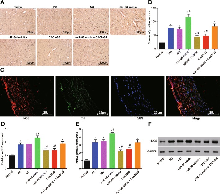
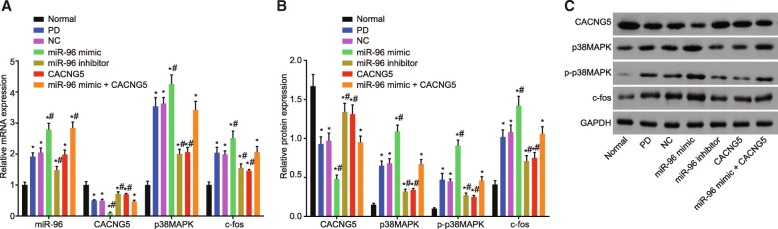
Background: There have been a number of reports implicating the association of microRNAs (miRs) and the MAPK signaling pathway with the dopaminergic neuron, which is involved in the development of Parkinson's disease (PD). The present study was conducted with aims of exploring the role of miR-96 in the activation of iNOS and apoptosis of dopaminergic neuron through the MAPK signaling pathway in mice with PD.

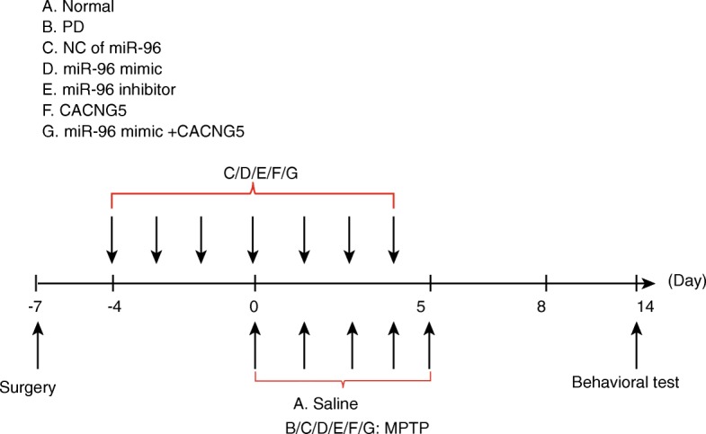
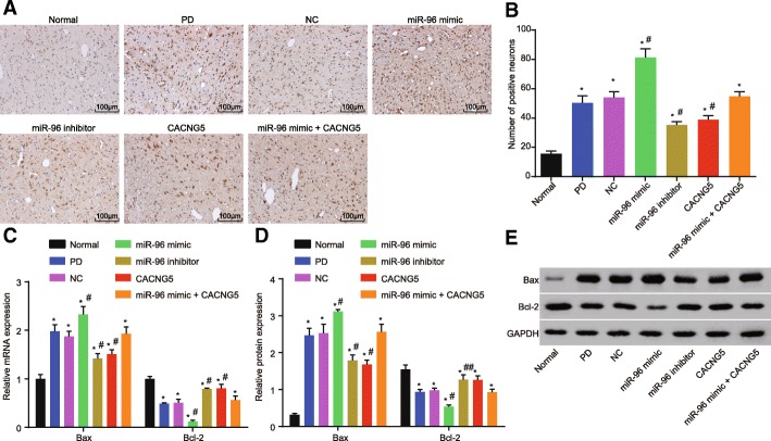
Methods: The miR and the differentially expressed gene in PD were screened out and the relationship between them was verified. A mouse model of PD induced by MPTP and was then constructed and treated with miR-96 mimic/inhibitor and CACNG5 overexpression plasmid to extract nigral dopaminergic neuron for the purpose of detecting the effect of miR-96 on PD. The TH and iNOS positive neuronal cells, the apoptotic neuronal cells by TUNEL staining, and expression of miR-96, CACNG5, iNOS, p38MAPK, p-p38MAPK, c-Fos, Bax, and Bcl-2 in substantia nigra dopaminergic neuronal tissues were evaluated.

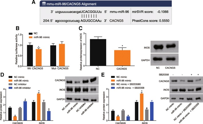
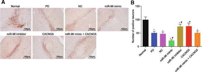
Results: The results obtained from the aforementioned procedure were then verified by cell culture of the SH-SY5Y cells, followed by treatment with miR-96 mimic/inhibitor, CACNG5 overexpression plasmid and the inhibitor of the MAPK signaling pathway. CACNG5 was confirmed as a target gene of miR-96. The inhibition of miR-96 resulted in a substantial increase in nigral cells, TH positive cells and expression of CACNG5 and Bcl-2 in nigral dopaminergic neuronal tissues, and a decrease in iNOS positive cells, apoptotic neuronal cells, and expression of iNOS, p38MAPK, p-p38MAPK, c-Fos, and Bax.

Conclusion: The above results implicated that the downregulation of miR-96 inhibits the activation of iNOS and apoptosis of dopaminergic neuron through the blockade of the MAPK signaling pathway by promoting CACNG5 in mice with PD.

Keywords: CACNG5; Dopaminergic neuron; Inducible nitric oxide synthase; MAPK signaling pathway; MicroRNA-96; Parkinson’s disease.


Conflict of interest statement

Ethics approval

The present study was performed in strict accordance with the ethics committee of China-Japan Union Hospital, Jilin University. All efforts were made to minimize the number of animals used and their suffering.

Consent for publication

Not applicable.

Competing interests

The authors declare that they have no competing interests.

Publisher’s Note

Springer Nature remains neutral with regard to jurisdictional claims in published maps and institutional affiliations.

Figures

Fig. 1
Fig. 1
The flowchart of model establishment and behavioral test. The horizontal axis represents the time points after the operation
Fig. 2
Fig. 2
miR-96 is predicted to regulate the MAPK signaling pathway via CACNG5. a prediction of target relationship between miR-96 and CACNG5 promoter region; b the effect of miR-96 on WT-CACNG5 and Mut-CACNG5 luciferase reporter plasmid; c the effect of CACNG5 on the expression of iNOS protein; d and e, effect of miR-96 on the expression of iNOS protein. *, P < 0.05 compared with the NC group. The test was repeated three times independently, and the non-paired t-test was used for comparison between the two groups; the one-way ANOVA analysis was used for comparison among multiple groups. miR-96, microRNA-96; CACNG5, calcium voltage-gated channel auxiliary subunit gamma 5; Wt, wild-type; Mut, mutant; NC, negative control
Fig. 3
Fig. 3
HE staining shows that miR-96 inhibitor increases the number of substantia nigra (× 100). n = 10 in each group; HE, hematoxylin-eosin; miR-96, microRNA-96; CACNG5, calcium voltage-gated channel auxiliary subunit gamma 5; PD, Parkinson’ disease; NC, negative control
Fig. 4
Fig. 4
miR-96 inhibitor increases expression of TH in substantia nigra dopaminergic neuronal tissues (× 100). a TH immunohistochemistry for dopaminergic neuronal tissues among seven groups; b positive TH cells among seven groups under a microscope; *, P < 0.05 compared with the normal group, #, P < 0.05 compared with the PD and NC groups. The test was repeated three times independently, and the one-way ANOVA analysis was used for comparison among multiple groups (n = 10 in each group). TH, tyrosine hydroxylase; miR-96, microRNA-96; CACNG5, calcium voltage-gated channel auxiliary subunit gamma 5; PD, Parkinson’ disease; NC, negative control
Fig. 5
Fig. 5
miR-96 inhibitor results in a decrease in the expression of iNOS in dopaminergic neuronal tissues. a iNOS immunohistochemistry for dopaminergic neuronal tissues among seven groups; b iNOS positive cells among seven groups under a microscope; c immunohistochemistry of colocalization analysis between the TH and iNOS; d mRNA expression of iNOS among seven groups evaluated by RT-qPCR; e protein expression of iNOS among seven groups; f protein bands of iNOS among seven groups evaluated by Western blot analysis; *, P < 0.05 compared with the normal group, #, P < 0.05 compared with the PD and NC groups; Bar = 50 μm. The test was repeated three times independently, and the one-way ANOVA analysis was used for comparison among multiple groups (n = 10 in each group). iNOS, inducible nitric oxide synthase; RT-qPCR, reverse transcription quantitative polymerase chain reaction; miR-96, microRNA-96; CACNG5, calcium voltage-gated channel auxiliary subunit gamma 5; PD, Parkinson’ disease; NC, negative control
Fig. 6
Fig. 6
Lower Bax and higher Bcl-2 were found after transfection of miR-96 inhibitor. a TUNEL staining of dopaminergic neuronal tissues among seven groups (× 100); b positive cells among seven groups under a microscope; c mRNA expression of Bax and Bcl-2 among seven groups evaluated by RT-qPCR; d protein expression of Bax and Bcl-2 among seven groups evaluated by Western blot; e protein bands of Bax and Bcl-2 among seven groups; Bar = 50 μm. The test was repeated three times independently, and the one-way ANOVA analysis was used for comparison among multiple groups (n = 10 in each group). *, P < 0.05 compared with the normal group, #, P < 0.05 compared with the PD and NC groups; TUNEL, terminal deoxynucleotidyl transferase-mediated dUTP nick-end-labeling; RT-qPCR, reverse transcription quantitative polymerase chain reaction; Bcl-2, B-cell lymphoma-2; Bax, Bcl-2 associated protein X; miR-96, microRNA-96; CACNG5, calcium voltage-gated channel auxiliary subunit gamma 5; PD, Parkinson’ disease; NC, negative control
Fig. 7
Fig. 7
Down-regulation of miR-96 leads to the inhibition of the MAPK signaling pathway through the up-regulation of CACNG5. a miR-96 expression and mRNA expression of CACNG5, p38MAPK and c-Fos evaluated by RT-qPCR among seven groups; b protein expression of CACNG5, p38MAPK, c-Fos, and p-p38MAPK evaluated by Western blot analysis among seven groups; c protein bands of CACNG5, p38MAPK, c-Fos, and p-p38MAPK; GAPDH used as a internal reference. The test was repeated three times independently, and the one-way ANOVA analysis was used for comparison among multiple groups (n = 10 in each group). *, P < 0.05 compared with the normal group, #, P < 0.05 compared with the PD and NC groups; miR-96, microRNA-96; CACNG5, calcium voltage-gated channel auxiliary subunit gamma 5; p38MAPK, p38 mitogen-activated protein kinase; RT-qPCR, reverse transcription quantitative polymerase chain reaction; PD, Parkinson’ disease; NC, negative control

Similar articles

Cited by

References

    1. Ayuk SM, Abrahamse H, Houreld NN. The role of photobiomodulation on gene expression of cell adhesion molecules in diabetic wounded fibroblasts in vitro. J Photochem Photobiol B. 2016;161:368–374. doi: 10.1016/j.jphotobiol.2016.05.027. - DOI - PubMed
    1. Brito MA, Vaz AR, Silva SL, Falcao AS, Fernandes A, Silva RF, et al. N-methyl-aspartate receptor and neuronal nitric oxide synthase activation mediate bilirubin-induced neurotoxicity. Mol Med. 2010;16:372–380. doi: 10.2119/molmed.2009-00152. - DOI - PMC - PubMed
    1. Chen C, Zhang X, Zhang S, Zhu S, Xu J, Zheng Y, et al. Quantification of protein copy number in single mitochondria: the Bcl-2 family proteins. Biosens Bioelectron. 2015;74:476–482. doi: 10.1016/j.bios.2015.06.057. - DOI - PubMed
    1. Crabtree DM, Zhang J. Genetically engineered mouse models of Parkinson’s disease. Brain Res Bull. 2012;88:13–32. doi: 10.1016/j.brainresbull.2011.07.019. - DOI - PMC - PubMed
    1. Dejager L, Vandevyver S, Petta I, Libert C. Dominance of the strongest: inflammatory cytokines versus glucocorticoids. Cytokine Growth Factor Rev. 2014;25:21–33. doi: 10.1016/j.cytogfr.2013.12.006. - DOI - PubMed