. 2018 Nov 28;18(1):178.
doi: 10.1186/s12862-018-1293-z.

Growth zone segmentation in the milkweed bug Oncopeltus fasciatus sheds light on the evolution of insect segmentation

Affiliations

Growth zone segmentation in the milkweed bug Oncopeltus fasciatus sheds light on the evolution of insect segmentation

Tzach Auman et al. BMC Evol Biol. .

Abstract

Background: One of the best studied developmental processes is the Drosophila segmentation cascade. However, this cascade is generally considered to be highly derived and unusual, with segments being patterned simultaneously, rather than the ancestral sequential segmentation mode. We present a detailed analysis of the segmentation cascade of the milkweed bug Oncopletus fasciatus, an insect with a more primitive segmentation mode, as a comparison to Drosophila, with the aim of reconstructing the evolution of insect segmentation modes.

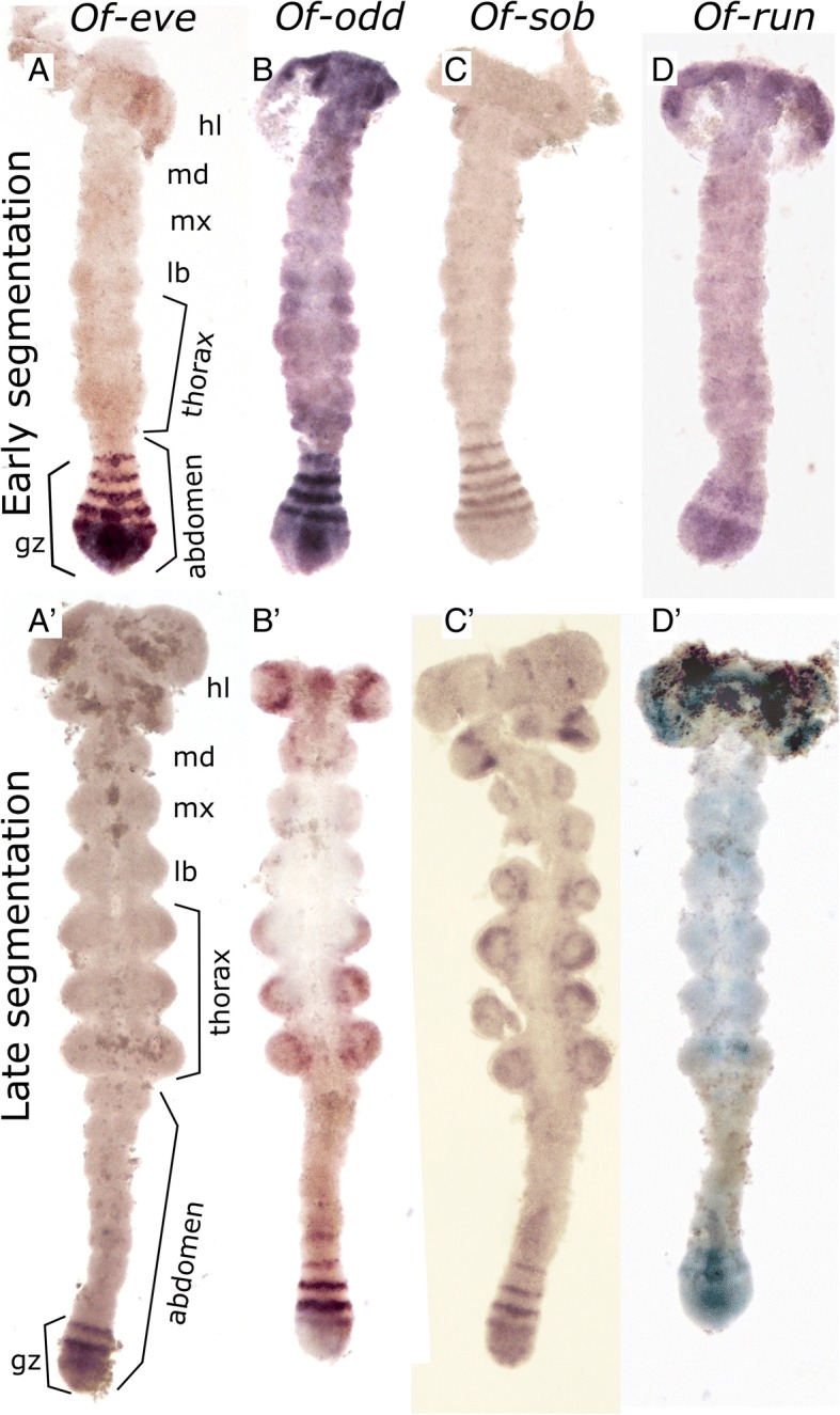
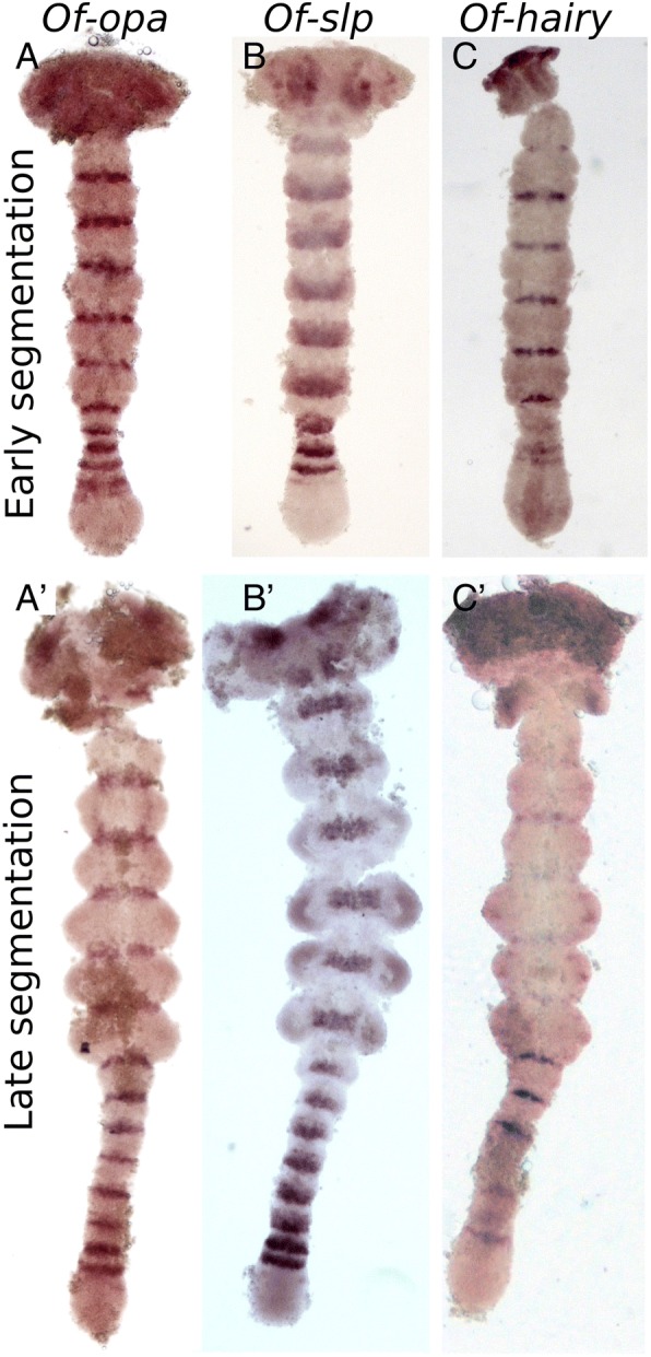
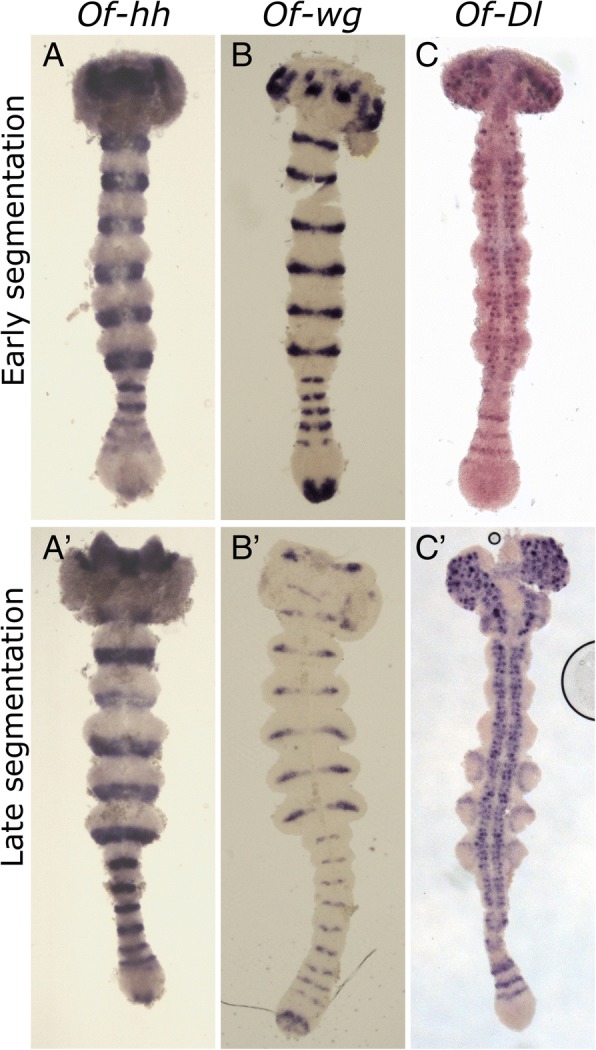
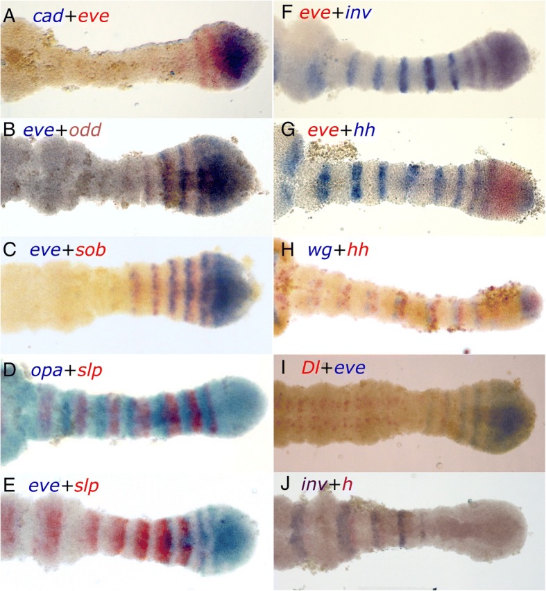
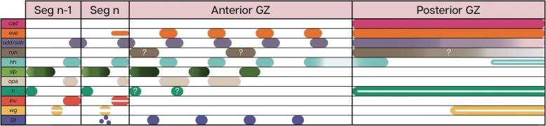
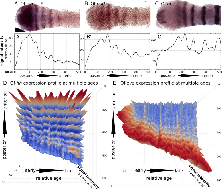
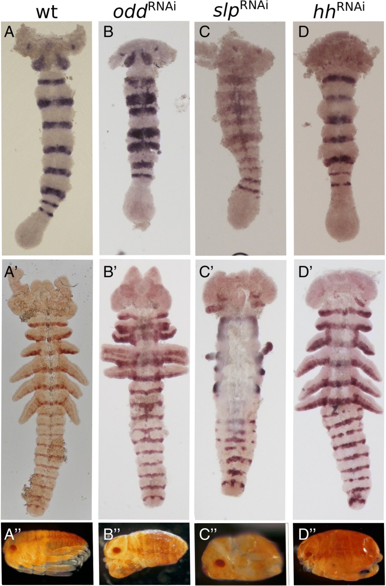
Results: We document the expression of 12 genes, representing different phases in the segmentation process. Using double staining we reconstruct the spatio-temporal relationships among these genes. We then show knock-down phenotypes of representative genes in order to uncover their roles and position in the cascade.

Conclusions: We conclude that sequential segmentation in the Oncopeltus germband includes three slightly overlapping phases: Primary pair-rule genes generate the first segmental gene expression in the anterior growth zone. This pattern is carried anteriorly by a series of secondary pair-rule genes, expressed in the transition between the growth zone and the segmented germband. Segment polarity genes are expressed in the segmented germband with conserved relationships. Unlike most holometabolous insects, this process generates a single-segment periodicity, and does not have a double-segment pattern at any stage. We suggest that the evolutionary transition to double-segment patterning lies in mutually exclusive expression patterns of secondary pair-rule genes. The fact that many aspects of the putative Oncopeltus segmentation network are similar to those of Drosophila, is consistent with a simple transition between sequential and simultaneous segmentation.

Keywords: Arthropod; Body plan; Evo-devo; Segment.


Conflict of interest statement

Ethics approval and consent to participate

All work reported herein was on invertebrate animals that do not require ethical approval.

Consent for publication

N/A

Competing interests

The authors declare they have no competing interests.

Publisher’s Note

Springer Nature remains neutral with regard to jurisdictional claims in published maps and institutional affiliations.

Figures

Fig. 1
Fig. 1
mRNA expression of the pair-rule genes eve (a-a’), odd (b, b′), sob (c-c′), and run (d-d’) in embryos at early and late abdominal segmentation. In the early segmenting germband (44–46 h after egg laying (hAEL)), eve (a), odd (b) and sob (c) all display a similar expression pattern in the anterior growth zone (GZ) composed of 4–6 stripes, corresponding to nascent segments. The main difference between the expression pattern of these genes is most notable in the posterior GZ where eve is steadily expressed, whereas odd and sob show weaker and graduated expression. Run (d-d’) expression is very different form the other three genes. It is expressed in two broad stripes corresponding to the anterior and posterior GZ, and in patches in the anterior thoracic and gnathal segments. In late germband stages (50–52 hAEL) (a’- d’) we see striped pattern of eve, odd and sob maintained, but with a smaller number of stripes. Expression of run is decreased to a single broad band in the anterior GZ. In addition, odd, sob and run are expressed in the limb buds. Embryos were chosen from within the aforementioned age range to be as similar as possible in developmental stage, based on size and shape of the germband and growth zone. In all images anterior is to the top. Abbreviations: gz, growth zone; hl, head lobe; md, mandibular segment; mx, maxillary segment; lb., labial segment
Fig. 2
Fig. 2
Expression of the pair rule genes opa (a-a’), slp (b-b′) and h (c-c’) in embryos at early and late abdominal segmentation. Throughout development, opa (a, a’) is expressed in a narrow band at the border of every segment but is not found in the posterior GZ. slp (b, b′) is more broadly and anteriorly expressed in each segment. The earliest, most posterior stripes are thin, and increase in breadth anteriorly. In later stages (b’), it shows diffuse expression in the limb buds. h expression (c-c′) is similar to that of opa in nascent segments but is weaker in mature segments. In the anterior GZ it is expressed in two stripes at the anterior of the anterior GZ, and more weakly in the posterior GZ. There is also weak punctate expression in the limb buds. Embryo ages are as in Fig. 1. In all images, anterior is to the top
Fig. 3
Fig. 3
Expression of the segment polarity genes hh (a-a’), and wg (b-b′), and the Notch ligand Dl (c-c). For the most part hh expression corresponds to that of inv/en, defining the posterior of each segment. Unlike inv/en, hh is expressed in the anterior GZ, and in a patch at the posterior GZ. wg (b, b′) is expressed in the middle of each segment. Like hh, it is expressed in a patch in the posterior of the GZ. Dl (c-c′) is found to be expressed in a series of stripes in the anterior GZ, and in a punctate pattern in neuronal tissue. Embryo ages are as in Fig. 1. In all images, anterior is to the top
Fig. 4
Fig. 4
Relative expression domains of different genes in the posterior of the embryo, as illustrated by combinations of double staining. a cad and eve are co-expressed in the posterior GZ, with eve stripes extending into the anterior GZ. b eve and odd, are shifted relative to each other, with overlapping expression in a narrow area (posterior of eve and anterior of odd), but with most of the expression separate. c The relationship between eve and sob is identical to that between eve and odd. d opa and slp are expressed in adjacent domains with no observable overlap. The posterior stripes are complementary and cover the entire anterior GZ. In later, more anterior stripes, as the segment grows, a region without opa or slp emerges, anterior to opa and posterior to slp. In later stages, opa and slp are completely separated. e slp expression begins just as eve expression is fading. In the segments where they are both expressed, slp is expressed to the anterior of eve. f The transition between eve and inv defines the GZ-germband border. At the transition, they are co-expressed in one or two stripes, in which their domains overlap. g The first stripes of hh expression overlap those of eve in the anterior GZ. h hh is immediately adjacent and posterior to wg in segmental stripes beginning in the anterior GZ. This relation is maintained in the posterior GZ, where both are expressed in non-overlapping patches. i Dl and eve are partially co-expressed in the anterior GZ, with Dl extending more anteriorly than eve, and eve beginning posteriorly to their overlapping domain. j h is expressed immediately posteriorly to inv, beginning slightly more anteriorly. Anterior is to the left in all images
Fig. 5
Fig. 5
Schematic representation of the relative expression patterns of all the genes discussed, as deduced from the double and single stainings. Question marks indicate cases with ambiguous staining, or where double staining was not possible, preventing us from identifying exact relative expression domains
Fig. 6
Fig. 6
Quantitative analysis of the dynamic expression of eve, odd and hh. a-c High magnification images of the growth zone and posterior germband of embryos stained for (a) eve (b) odd and (c) hh. (a’-c′) Gene expression levels in the embryo shown above. For each embryo, we drew a rectangle encompassing the entire imaged region and summed the pixel intensities for each point along the x-axis (posterior to anterior). Comparison of the signal intensity highlights the small differences in the expression profile of these genes in the GZ. The main difference seems to be that while eve is robustly expressed in the posterior GZ and is strongest in the anterior of the posterior GZ, odd is weakly expressed in the posterior GZ, increasing in strength towards the anterior, and peaking only in the first discrete odd stripe in the anterior GZ. The main observation regarding the hh expression profile is the double-peak between the stripe in the anterior GZ and first stripe of the posterior GZ, which are not completely resolved. Only the third hh stripe is completely resolved. d-e 3D plots including a sequence of embryos expressing (d) hh and (e) eve, arranged in sequence by increasing total length of the GZ + abdominal segments. The third thoracic segment (T3) was defined as the zero point of the y-axis of each graph. For eve n = 71 for hh n = 53
Fig. 7
Fig. 7
Segmental phenotypes following knock-down of odd (b-b″), slp (c-c″) and hh (d-d”), in early and late germband embryos stained for inv and in hatchlings. a-a” Wildtype embryos and hatchling. b In the early germband embryo odd RNAi embryos mainly display widening of inv expression in the thoracic segments, and fusion of segments in the embryonic midline. b′ In later stages, appendages are fused, and the borders of some abdominal segments are also ill-defined, sporadically fused or narrowed. In both embryonic stages, slight ectopic expression of Of-inv is seen in single cells. b″ In the odd-RNAi hatchling this phenotype causes compression of the thorax and truncated limbs. c in slp-RNAi embryos thoracic inv expression is broader in the early germband embryo, and abnormally expressed in the midline. c′ The later slp-RNAi embryo displays severe truncation of all appendages, with only limb buds of T1 and T3 remaining. In addition, we see malformation of the abdominal segment boundaries, where gaps in inv expression can be seen. The slp-RNAi embryo is also wider than WT embryos and has an apparent breakdown of midline tissues. c″ The slp-RNAi hatchlings are compressed with almost no segmental boundaries, and holes appear in the lateral parts of the embryo, where the limbs are missing. d Early hh-RNAi embryos seem to be almost completely normal, only displaying some minor head aberration. d’ Aberrations of the head are also seen in the late germband embryos which seems to lack some folds and finer details of the head structure. Abdominal segment borders are also affected, containing gaps and ectopic expression of inv in sporadic cells. d” In hatchlings, the head is greatly reduced and malformed. Segmental borders can be seen, but they are disrupted. Limbs develop normally

Similar articles

Cited by

References

    1. Peel AD, Chipman AD, Akam M. Arthropod segmentation: beyond the Drosophila paradigm. Nat Rev Genet. 2005;6(12):905–916. doi: 10.1038/nrg1724. - DOI - PubMed
    1. Lawrence PA. The making of a fly: the genetics of animal design. Oxford: Blackwell Scientific Publications; 1992.
    1. Hartenstein V, Chipman AD. Hexapoda: a Drosophila’s view of insect development. In: Wanninger A, editor. Evolutionary Developmental Biology of Invertebrates. Vienna: Springer; 2015. pp. 1–91.
    1. Stahi R, Chipman AD. Blastoderm segmentation in Oncopeltus fasciatus and the evolution of arthropod segmentation mechanisms. Proc R Soc Lond B. 2016;283:20161745. doi: 10.1098/rspb.2016.1745. - DOI - PMC - PubMed
    1. Peel A. The evolution of arthropod segmentation mechanisms. Bioessays. 2004;26(10):1108–1116. doi: 10.1002/bies.20097. - DOI - PubMed

Publication types

LinkOut - more resources