. 2018 Nov 29;20(1):264.
doi: 10.1186/s13075-018-1752-3.

The presence of anti-nuclear antibodies alone is associated with changes in B cell activation and T follicular helper cells similar to those in systemic autoimmune rheumatic disease

Affiliations

The presence of anti-nuclear antibodies alone is associated with changes in B cell activation and T follicular helper cells similar to those in systemic autoimmune rheumatic disease

Yuriy Baglaenko et al. Arthritis Res Ther. .

Abstract

Background: Diagnosis of systemic autoimmune rheumatic diseases (SARD) relies on the presence of hallmark anti-nuclear antibodies (ANA), many of which can be detected years before clinical manifestations. However, ANAs are also seen in healthy individuals, most of whom will not develop SARD. Here, we examined a unique cohort of asymptomatic ANA+ individuals to determine whether they share any of the cellular immunologic features seen in SARD.

Methods: Healthy ANA- controls and ANA+ (ANA ≥1:160 by immunofluorescence) participants with no SARD criteria, with at least one criterion (undifferentiated connective tissue disease (UCTD)), or meeting SARD classification criteria were recruited. Peripheral blood cellular immunological changes were assessed by flow cytometry and transcript levels of BAFF, interferon (IFN)-induced and plasma cell-expressed genes were quantified by NanoString.

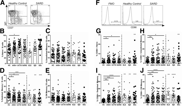
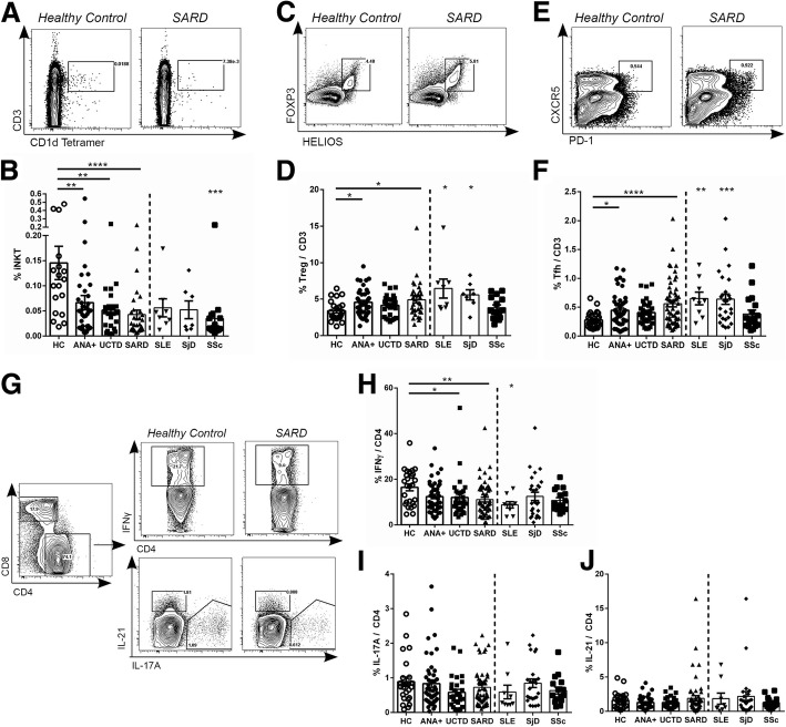
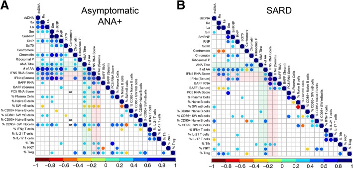
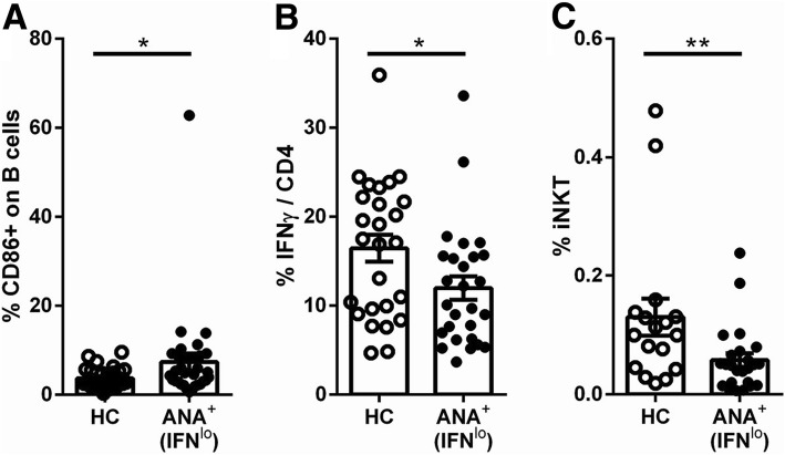
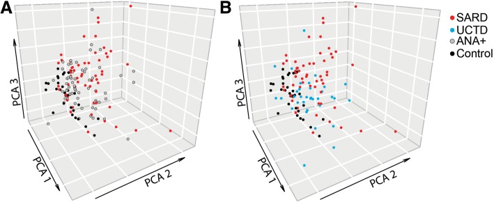
Results: A number of the immunologic abnormalities seen in SARD, including changes in peripheral B (switched memory) and T (iNKT, T regulatory, activated memory T follicular helper) subsets and B cell activation, were also seen in asymptomatic ANA+ subjects and those with UCTD. The extent of these immunologic changes correlated with ANA titer or the number of different specific ANAs produced. Principal component analysis of the cellular data indicated that a significant proportion of asymptomatic ANA+ subjects and subjects with UCTD clustered with patients with early SARD, rather than ANA- healthy controls.

Conclusions: ANA production is associated with altered T and B cell activation even in asymptomatic individuals. Some of the currently accepted cellular features of SARD may be associated with ANA production rather than the immunologic events that cause symptoms in SARD.

Keywords: Anti-nuclear antibodies; B cell; Systemic autoimmune rheumatic disease; T cell.


Conflict of interest statement

Ethics approval and consent to participate

The study was approved by the Research Ethics Boards of the University Health Network (12-5455-BE) and Mount Sinai Hospital, and all participants signed informed consent.

Consent for publication

Not applicable.

Competing interests

The authors declare that they have no competing interests.

Publisher’s Note

Springer Nature remains neutral with regard to jurisdictional claims in published maps and institutional affiliations.

Figures

Fig. 1
Fig. 1
Asymptomatic anti-nuclear antibodies (ANA)+ individuals with no diagnosis of systemic autoimmune rheumatic disease (SARD) have abnormalities in peripheral B cell subsets and their activation similar to symptomatic patients with SARD. a Gating strategy for identification of naïve (IgD+CD27), unswitched memory (IgD+CD27+), switched memory (IgDCD27+), and double-negative (DN) memory (IgDCD27) cells within the CD19+ B cell compartment of peripheral blood mononuclear cells from a representative healthy control and a patient with SARD. b–e Proportions of the different peripheral B cell subsets within the CD19+ B cell compartment for each subject group. f Representative gating for CD86+ cells within the various B cell compartments (example shown is gated on all CD19+ B cells). g–j Proportion of CD86+ cells within each of the different peripheral B cell subsets. Statistical comparisons on the left side of each plot are between healthy controls (HC) and asymptomatic ANA+ individuals (ANA+), patients with undifferentiated connective tissue disease (UCTD), or pooled patients with SARD, whereas those on the right side of the plot are comparisons between the individual SARDs and HC. Bars represent the mean with SEM. Every data point represents an individual patient. For each set of comparisons statistical significance was determined using the Kruskal-Wallis test with Dunn’s post-hoc test for multiple comparisons, as compared to HC; *p ≤ 0.05, **p ≤ 0.01, ***p ≤ 0.001, ****p ≤ 0.0001. SLE, systemic lupus erythematosus; SjD, Sjogren’s disease; SSc, systemic sclerosis
Fig. 2
Fig. 2
Reductions in invariant natural killer T (iNKT) cells and increases in T follicular helper (Tfh) and T regulatory (Treg) cells in anti-nuclear antibodies (ANA)+ individuals regardless of diagnosis. Representative gating, together with scatterplots show frequencies in each subject group for peripheral iNKT (a, b), Treg (c, d), and Tfh (e, f) cells. For the iNKT cell plot, the cells were first gated on lymphocytes, whereas for the Tfh and Treg cell plots, they were first gated on CD3+CD4+ cells within the lymphocyte pool with the scatterplots showing the percentage of these cells as a proportion of the CD3+ T cell population. g Representative gating for interferon (IFN)-γ-, IL-17A-, and IL-21-producing CD4 T cells following phorbol-12-myristate-13-acetate (PMA) and ionomycin stimulation of peripheral blood mononuclear cells (plots shown have been gated on CD3+CD4+ lymphocytes). h–j Proportions of cytokine-producing cells within the CD3+CD4+ T cell population for each subject group. Statistical comparisons on the left side of each figure are between healthy controls (HC) and asymptomatic ANA+ individuals (ANA+), patients with undifferentiated connective tissue disease (UCTD), or pooled patients with systemic autoimmune rheumatic disease (SARD), whereas those on the right side of the figure are between the individual SARDs and HC. Bars represent the mean with SEM. Every data point represents an individual patient. Statistical significance was determined using the Kruskal-Wallis test with Dunn’s post-hoc test for multiple comparisons, as compared to HC; *p ≤ 0.05, **p ≤ 0.01, ***p ≤ 0.001, ****p ≤ 0.0001. SLE, systemic lupus erythematosus; SjD, Sjogren’s disease; SSc, systemic sclerosis
Fig. 3
Fig. 3
Correlation between cellular and selected serologic/cytokine phenotypes in asymptomatic anti-nuclear antibodies (ANA)+ individuals and patients with systemic autoimmune rheumatic disease (SARD). Spearman correlation matrices for asymptomatic ANA+ individuals (a) and patients with SARD (b). B and T cell populations were gated and defined as outlined in Figs. 1 and 2, plasma cells as outlined in Additional file 1: Figure S3, and CD95+ B cell subpopulations as shown in Additional file 1: Figure S1. The color and size of the dots represents the ρ value, with the scales shown at the bottom of each matrix. Non-significant (p ≥ 0.05) correlation is not displayed. IFN, interferon; BAFF, B cell activating factor
Fig. 4
Fig. 4
Cellular immunologic changes seen in asymptomatic anti-nuclear antibodies (ANA)+ individuals who do not have elevations of type I interferons (IFNs). Asymptomatic ANA+ individuals were defined as having normal levels of type I IFNs (IFNlo) if their IFN5 score was < 2 SD above the mean for ANA healthy controls (HC) (all had undetectable levels of serum IFN-α on ELISA). Shown are cellular phenotypes (a % CD86+ B cells (CD86+ on B cells), b % CD4 cells producing IFN-γ (IFNγ/CD4) and c % invariant natural killer T cells (iNKT))  that were significantly different in IFNlo asymptomatic ANA+ individuals as compared to HC. Cell populations were defined and gated as outlined in Figs. 1 and 2. Bars represent the mean with SEM. Every data point represents an individual patient. Statistical significance was determined using the Mann-Whitney U test; *p ≤ 0.05, **p ≤ 0.01
Fig. 5
Fig. 5
In principal component analysis (PCA) the cellular phenotype of many asymptomatic anti-nuclear antibody (ANA)+ individuals or patients with undifferentiated connective tissue disease (UCTD) overlaps with that of patients with systemic autoimmune rheumatic disease (SARD) and is distinct from that of healthy controls. The 3-D plots show asymptomatic ANA+ individuals (ANA+, gray) (a) or patients with UCTD (blue) (b), as compared to healthy controls (black) and patients with SARD (red). Analyses were performed using the PCA function in the missMDA (v1.12) package, with missing data imputed using the imputePCA function. Corresponding plots were created using the scatterplot3d (v0.3–41) package

Similar articles

Cited by

References

    1. Alarcon-Segovia D, Alarcon-Riquelme ME, Cardiel MH, Caeiro F, Massardo L, Villa AR, Pons-Estel BA. Familial aggregation of systemic lupus erythematosus, rheumatoid arthritis, and other autoimmune diseases in 1,177 lupus patients from the GLADEL cohort. Arthritis Rheum. 2005;52(4):1138–1147. doi: 10.1002/art.20999. - DOI - PubMed
    1. Kallenberg CG. Overlapping syndromes, undifferentiated connective tissue disease, and other fibrosing conditions. Curr Opin Rheumatol. 1995;7(6):568–573. doi: 10.1097/00002281-199511000-00017. - DOI - PubMed
    1. Reveille JD, Wilson RW, Provost TT, Bias WB, Arnett FC. Primary Sjogren’s syndrome and other autoimmune diseases in families. Prevalence and immunogenetic studies in six kindreds. Ann Intern Med. 1984;101(6):748–756. doi: 10.7326/0003-4819-101-6-748. - DOI - PubMed
    1. Rodriguez-Reyna TS, Alarcon-Segovia D. Overlap syndromes in the context of shared autoimmunity. Autoimmunity. 2005;38(3):219–223. doi: 10.1080/08916930500050145. - DOI - PubMed
    1. Arbuckle MR, McClain MT, Rubertone MV, Scofield RH, Dennis GJ, James JA, Harley JB. Development of autoantibodies before the clinical onset of systemic lupus erythematosus. N Engl J Med. 2003;349(16):1526–1533. doi: 10.1056/NEJMoa021933. - DOI - PubMed

Publication types

MeSH terms

Substances

Grants and funding