

miR-663a inhibits tumor growth and invasion by regulating TGF-β1 in hepatocellular carcinoma
- PMID: 30486878
- PMCID: PMC6264054
- DOI: 10.1186/s12885-018-5016-z
miR-663a inhibits tumor growth and invasion by regulating TGF-β1 in hepatocellular carcinoma
Abstract
Background: The dysregulation of miR-663a is frequently observed in many human cancers. However, the functional role and precise mechanism of miR-663a have been controversial in hepatocellular carcinoma (HCC) and need to be studied in depth.
Methods: The expression of miR-663a was detected in human cell lines and HCC tissues by quantitative RT-PCR (qRT-PCR), and data from the Cancer Genome Atlas (TCGA). Cell proliferation was investigated using MTS, EdU, colony formation assays, and xenograft animal experiments, and the cell invasion capacity was evaluated using the transwell assay. The target gene of miR-663a was identified by qRT-PCR, Western blot, and dual-luciferase reporter assays. The clinicopathological features of miR-663a and the correlation between miR-663a and TGF-β1 expression were also investigated in the clinical samples of HCC.
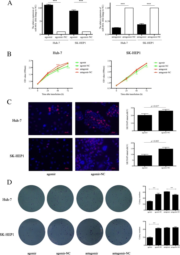
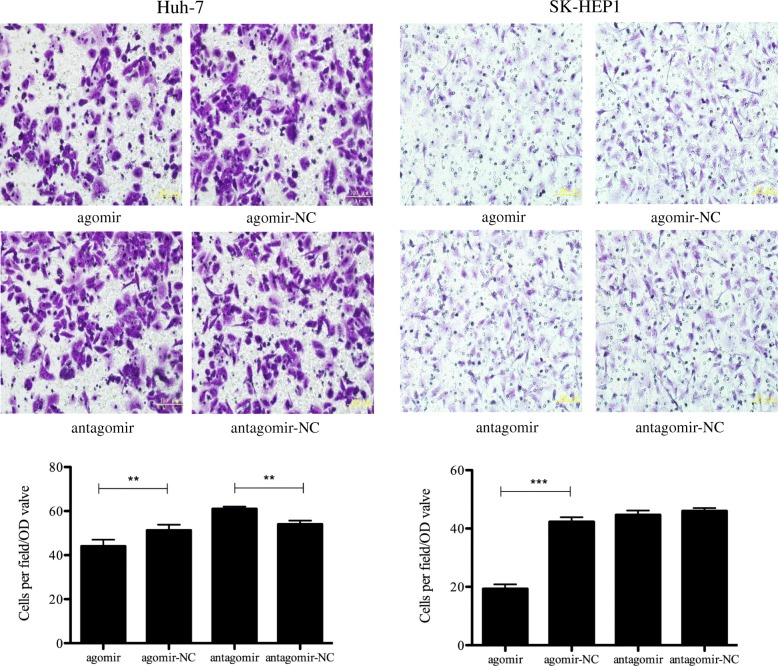
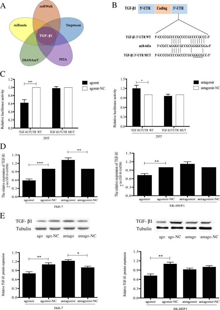
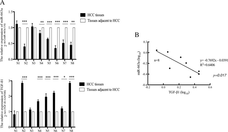
Results: miR-663a was significantly downregulated in HCC cells relative to immortal normal liver cells, as indicated using qRT-PCR, and the lower expression of miR-663a was also confirmed in HCC tissue samples and the data from TCGA. The expression of miR-663a in HCC tissue samples was statistically significantly associated with size and the number of tumors. In addition, the upregulation of miR-663a inhibited the proliferation and invasion of HCC cells in vitro. Further study showed that miR-663a directly targeted transforming growth factor beta 1 (TGF-β1) to suppress HCC invasion, and that the inhibitory effect of miR-663a on cell invasion could be regulated by TGF-β1. In vivo studies showed that miR-663a significantly inhibited tumor growth. A negative correlation between miR-663a and TGF-β1 expression was also confirmed from the clinical samples of HCC.
Conclusions: miR-663a acts as a tumor suppressor and exerts a substantial role in inhibiting the proliferation, invasion, and tumorigenesis of HCC by regulating TGF-β1 in vitro and in vivo. These observations indicate that miR-663a may be a suitable diagnostic, therapeutic, and prognostic target for the treatment of HCC.
Keywords: Hepatocellular carcinoma (HCC); Invasion; Proliferation; Transforming growth factor β1 (TGF-β1); miR-663a.
Conflict of interest statement
Ethics approval and consent to participate
The study protocol was approved by the Institutional Research Ethics Committee of the First Hospital of China Medical University. The informed consent to participate in the study from the patients was also written. For in vivo studies on animals, the study was also approved by the Institutional Research Ethics Committee of the hospital in accordance with the Guide for the Care and Use of Laboratory Animals. The human cell lines did not require ethics approval from our hospital as they were purchased from legal commercial product.
Consent for publication
Not applicable.
Competing interests
The authors declare that they have no competing interests.
Publisher’s Note
Springer Nature remains neutral with regard to jurisdictional claims in published maps and institutional affiliations.
Figures


Similar articles
-
Downregulation of lncRNA SBF2-AS1 inhibits hepatocellular carcinoma proliferation and migration by regulating the miR-361-5p/TGF-β1 signaling pathway.Aging (Albany NY). 2021 Aug 2;13(15):19260-19271. doi: 10.18632/aging.203248. Epub 2021 Aug 2. Aging (Albany NY). 2021. PMID: 34341185 Free PMC article.
-
miR-663a inhibits hepatocellular carcinoma cell proliferation and invasion by targeting HMGA2.Biomed Pharmacother. 2016 Jul;81:431-438. doi: 10.1016/j.biopha.2016.04.034. Epub 2016 Apr 28. Biomed Pharmacother. 2016. PMID: 27261623
-
NEAT1 upregulates TGF-β1 to induce hepatocellular carcinoma progression by sponging hsa-mir-139-5p.J Cell Physiol. 2018 Nov;233(11):8578-8587. doi: 10.1002/jcp.26524. Epub 2018 May 24. J Cell Physiol. 2018. PMID: 29797561
-
The dichotomous role of TGF-β in controlling liver cancer cell survival and proliferation.J Genet Genomics. 2020 Sep 20;47(9):497-512. doi: 10.1016/j.jgg.2020.09.005. Epub 2020 Nov 14. J Genet Genomics. 2020. PMID: 33339765 Review.
-
Antitumor function of microRNA-122 against hepatocellular carcinoma.J Gastroenterol. 2014 Apr;49(4):589-93. doi: 10.1007/s00535-014-0932-4. Epub 2014 Feb 17. J Gastroenterol. 2014. PMID: 24531873 Review.
Cited by
-
Comprehensive profiling and characterization of cellular microRNAs in response to coxsackievirus A10 infection in bronchial epithelial cells.Virol J. 2022 Jul 21;19(1):120. doi: 10.1186/s12985-022-01852-9. Virol J. 2022. PMID: 35864512 Free PMC article.
-
Novel DNA methylation-based epigenetic signatures in colorectal cancer from peripheral blood leukocytes.Am J Cancer Res. 2024 May 15;14(5):2253-2271. doi: 10.62347/MXWJ1398. eCollection 2024. Am J Cancer Res. 2024. PMID: 38859857 Free PMC article.
-
LncRNA miR663AHG represses the development of colon cancer in a miR663a-dependent manner.Cell Death Discov. 2023 Jul 3;9(1):220. doi: 10.1038/s41420-023-01510-1. Cell Death Discov. 2023. PMID: 37400477 Free PMC article.
-
Epigenetic strategies to boost CAR T cell therapy.Mol Ther. 2021 Sep 1;29(9):2640-2659. doi: 10.1016/j.ymthe.2021.08.003. Epub 2021 Aug 6. Mol Ther. 2021. PMID: 34365035 Free PMC article. Review.
-
Irradiation-responsive PRDM10-DT modulates the angiogenic response in human NSCLC cells in an SP1-dependent manner via the miR-663a/TGF-β1 axis.J Transl Med. 2025 Feb 27;23(1):235. doi: 10.1186/s12967-025-06273-0. J Transl Med. 2025. PMID: 40016776 Free PMC article.
References
-
- Zhang CS, Zhang JL, Li XH, Li L, Li X, Zhou XY. Is radiofrequency ablation equal to surgical re-resection for recurrent hepatocellular carcinoma meeting the Milan criteria? A meta-analysis. J BUON. 2015;20(1):223–230. - PubMed
MeSH terms
Substances
Grants and funding
LinkOut - more resources
Full Text Sources
Medical
Miscellaneous
