. 2018 Nov 28;37(1):291.
doi: 10.1186/s13046-018-0972-3.

Hypoxia potentiates gemcitabine-induced stemness in pancreatic cancer cells through AKT/Notch1 signaling

Affiliations

Hypoxia potentiates gemcitabine-induced stemness in pancreatic cancer cells through AKT/Notch1 signaling

Zhengle Zhang et al. J Exp Clin Cancer Res. .

Abstract

Background: Profound chemoresistance remains an intractable obstacle in pancreatic cancer treatment. Pancreatic cancer stem cells (CSCs) and the ubiquitous hypoxic niche have been proposed to account for drug resistance. However, the mechanism involved requires further exploration. This study investigated whether the hypoxic niche enhances gemcitabine-induced stemness and acquired resistance in pancreatic cancer cells by activating the AKT/Notch1 signaling cascade. The therapeutic effects of blockading this signaling cascade on gemcitabine-enriched CSCs were also investigated.

Methods: The expression levels of CSC-associated markers Bmi1 and Sox2 as well as those of proteins involved in AKT/Notch1 signaling were measured by Western blot analysis. The expression level of the pancreatic CSC marker CD24 was measured by flow cytometry. Change in gemcitabine sensitivity was evaluated by the MTT assay. The ability of sphere formation was tested by the sphere-forming assay in stem cell medium. The ability of migration and invasion was detected by the transwell migration/invasion assay. A mouse xenograft model of pancreatic cancer was established to determine the effect of Notch1 inhibition on the killing effect of gemcitabine in vivo. The ability of metastasis was investigated by an in vivo lung metastasis assay.

Results: Gemcitabine promoted pancreatic cancer cell stemness and associated malignant phenotypes such as enhanced migration, invasion, metastasis, and chemoresistance. The AKT/Notch1 signaling cascade was activated after gemcitabine treatment and mediated this process. Blockading this pathway enhanced the killing effect of gemcitabine in vivo. However, supplementation with hypoxia treatment synergistically enhanced the AKT/Notch1 signaling pathway and collaboratively promoted gemcitabine-induced stemness.

Conclusions: These findings demonstrate a novel mechanism of acquired gemcitabine resistance in pancreatic cancer cells through induction of stemness, which was mediated by the activation of AKT/Notch1 signaling and synergistically aggravated by the ubiquitous hypoxic niche. Our results might provide new insights for identifying potential targets for reversing chemoresistance in patients with pancreatic cancer.

Keywords: AKT; Cancer stem cell; Gemcitabine; Hypoxia; Notch1.


Conflict of interest statement

Ethics approval and consent to participate

This study was approved by the ethical review board of Renmin Hospital, Wuhan University (Wuhan, China).

Consent for publication

Not applicable.

Competing interests

The authors declare that they have no competing interests.

Publisher’s Note

Springer Nature remains neutral with regard to jurisdictional claims in published maps and institutional affiliations.

Figures

Fig. 1
Fig. 1
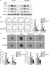
Gemcitabine promotes Notch1 activation and pancreatic cancer cell stemness. (a) PANC-1 and Patu8988 cells were treated with 0.1–500 μM gemcitabine for 24 h, and the relative survival rate was measured by the MTT assay. Western blot findings revealed (b) the representative expression levels of Bmi1, Sox2, NICD1, and Notch1 as well as (c) the changes in these levels after treatment with different concentrations of gemcitabine for 24 h. After treatment with 5 μM gemcitabine for 24 h, (d) the representative expression level of the pancreatic CSC marker CD24 as well as (e) the change in the proportion of CD24+ pancreatic CSCs were determined by FCM. (f-h) The ability of the cells to form spheres after treatment was evaluated by the sphere-forming assay in stem cell medium: (f) Representative image of sphere formation in cancer cells; (g, h) Charts showing the data on sphere number and diameter. The data are derived from three independent assays. Scale bar, 50 μm. *P < 0.05; **P < 0.01; ***P < 0.001
Fig. 2
Fig. 2
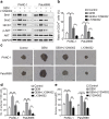
Notch1 signaling mediates gemcitabine-induced stemness. PANC-1 and Patu8988 cells were pretreated with 10 μM DAPT for 24 h and then treated with gemcitabine. (a) The expression levels of Bmi1, Sox2, and NICD1 were determined by Western blot analysis. (b) The representative expression level of the pancreatic CSC marker CD24 as well as (c) the change in the proportion of CD24+ pancreatic CSCs were determined by FCM. (d-f) The ability of the cells for sphere formation after treatment was determined by the sphere-forming assay: (d) Representative image of spheres formed after treatment; (e, f) Charts showing the data on sphere number and size. The results presented are from three independent assays. Scale bar, 50 μm. *P < 0.05; **P < 0.01; ***P < 0.001
Fig. 3
Fig. 3
Notch1 inhibition enhances the killing effect of gemcitabine in vivo. PANC-1 cells were subcutaneously injected into the right flank of nude mice. After about 6 days, the mice were randomly divided into the control, DAPT, GEM, and GEM+DAPT groups in accordance with the protocol described in the Methods. (a) Representative tumor size at 38 days post-treatment. (b) Tumor growth curves delineated on the basis of volume measured every 4 days. (c) After treatment, the expression levels of Bmi1, Sox2, NICD1, and Notch1 were determined by Western blot analysis. (d, e) Tumor samples were digested by using collagenase I, and the change in the proportion of CD24+ pancreatic CSCs was determined by FCM. The graphs are from three independent experiments. **P < 0.01
Fig. 4
Fig. 4
AKT promotes pancreatic cancer cell stemness partly by mediating Notch1 activation. Two cell lines were pretreated with 20 μM LY294002 for 2 h and then treated with gemcitabine. (a) The expression levels of Bmi1, Sox2, NICD1, p-AKT (serine 473), and AKT were determined by Western blot analysis. (b) The change in the proportion of CD24+ pancreatic CSCs was determined by FCM. (c-e) The ability of the cells for sphere formation was investigated by the sphere-forming assay: (c) Representative image of spheres formed after treatment; (d and e) Charts showing the data on sphere number and size. The graphs show the results of three independent experiments. Scale bar, 50 μm. *P < 0.05; **P < 0.01; ***P < 0.001
Fig. 5
Fig. 5
Hypoxia synergistically enhances gemcitabine-induced stemness. (a) Two pancreatic cancer cell lines were incubated under different hypoxic (1%) conditions, and the expression levels of Bmi1, Sox2, and HIF-1α were determined by Western blot analysis. (b) After treatment with different concentrations of CoCl2 for 24 h, the changes in Bmi1, Sox2, and HIF-1α expression levels were determined by Western blot analysis. (c) Two cell lines were co-treated with gemcitabine and CoCl2 for 24 h, and the protein expression levels were measured by Western blot analysis. (d) The sphere formation ability of the cells after co-treatment was determined by the sphere-forming assay. (e, f) Charts showing the data on sphere number and size after treatment. The graphs are from three independent experiments. Scale bar, 50 μm. *P < 0.05; **P < 0.01
Fig. 6
Fig. 6
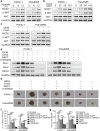
AKT/Notch1 signaling mediates the synergistic enhancement of stemness induced by gemcitabine and hypoxia. (a) PANC-1 and Patu8988 cells were treated under hypoxic conditions (1%) for different durations, and the expression levels of NICD1, p-AKT, and AKT were determined by Western blot analysis. (b) After treatment with different concentrations of CoCl2, the changes in NICD1, p-AKT, and AKT expression levels were determined by Western blot analysis. (c) Two cell lines were co-treated with gemcitabine and CoCl2, and the protein expression levels were determined by Western blot analysis. (d) Two pancreatic cancer cell lines were pretreated with 10 μM DAPT or 20 μM LY294002 and then treated with a combination of gemcitabine and CoCl2. Then, the expression levels of Bmi1, Sox2, and NICD1 were determined by Western blot analysis. (e-g) After this treatment, the two cell lines were cultured with stem cell medium, and the ability of the cells for sphere formation was investigated by the sphere-forming assay: (e) Representative image of spheres after treatment; (f, g) Charts showing the data on sphere number and size. All data shown are from three independent experiments. Scale bar, 50 μm. *P < 0.05; **P < 0.01; ***P < 0.001
Fig. 7
Fig. 7
Schematic illustration demonstrating how hypoxia potentiates gemcitabine-induced stemness and acquired resistance in pancreatic cancer cells through AKT/Notch1 signaling

Similar articles

Cited by

References

    1. Siege RL, Miller KD, Jemal A. Cancer statistics, 2016. CA Cancer J Clin. 2016;66:7–30. doi: 10.3322/caac.21332. - DOI - PubMed
    1. Ryan DP, Hong TS, Bardeesy N. Pancreatic adenocarcinoma. N Engl J Med. 2014;371:2140–2141. doi: 10.1056/NEJMra1404198. - DOI - PubMed
    1. Clevers H. The cancer stem cell: premises, promises and challenges. Nat Med. 2011;17:313–319. doi: 10.1038/nm.2304. - DOI - PubMed
    1. Bergman AM, Pinedo HM, Peters GJ. Determinants of resistance to 2′,2′-difluorodeoxycytidine (gemcitabine) Drug Resist Updat. 2002;5:19–33. doi: 10.1016/S1368-7646(02)00002-X. - DOI - PubMed
    1. Ansari D, Tingstedt B, Andersson B, Holmquist F, Sturesson C, Williamsson C, et al. Pancreatic cancer: yesterday, today and tomorrow. Future Oncol. 2016;12:1929–1946. doi: 10.2217/fon-2016-0010. - DOI - PubMed

MeSH terms