. 2018 Nov 28;49(1):116.
doi: 10.1186/s13567-018-0611-1.

Interspecies transmission to bovinized transgenic mice uncovers new features of a CH1641-like scrapie isolate

Affiliations

Interspecies transmission to bovinized transgenic mice uncovers new features of a CH1641-like scrapie isolate

Kohtaro Miyazawa et al. Vet Res. .

Abstract

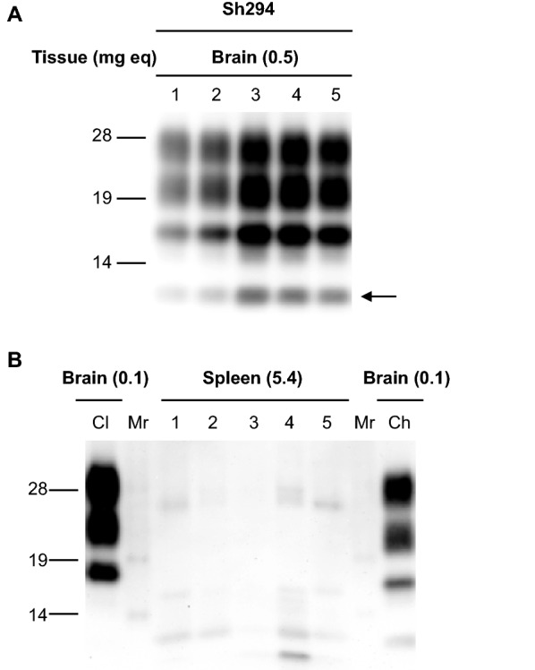
In animal prion diseases, including bovine spongiform encephalopathy (BSE) in cattle, chronic wasting disease in cervids, and scrapie in sheep and goats, a disease-associated isoform of prion protein (PrPd) accumulates in the brains of affected animals. Although the CH1641 scrapie isolate was experimentally established in the UK, a few natural CH1641-like scrapie cases have been reported in France and the UK. The molecular mass of the unglycosylated protease-resistant core of PrPd (PrPres) is known to be similar between CH1641-like scrapie and experimental BSE in sheep. We previously established an experimental CH1641-like scrapie isolate (Sh294) from a natural classical scrapie case. Here, we demonstrated that the Sh294 isolate was independent of both classical and atypical BSEs by cross-species transmission to bovine PrP overexpressing (TgBoPrP) mice and wild-type mice. Interestingly, we found that the Sh294 isolate altered its host range by the transmission to TgBoPrP mice, and we succeeded in the first stable reproduction of CH1641-like scrapie specific PrPres banding patterns with the ~12-kDa small C-terminal fragment in wild-type mice. This study provides new insight into the relationship between CH1641-like scrapie isolates and BSEs. In addition, interspecies transmission models such as we have demonstrated here could be a great help to investigate the origin and host range of animal prions.


Figures

Figure 1
Figure 1
Western blot analysis of TgOvPrP59 mice infected with Sh294. Brains and spleens were dissected from Sh294-infected TgOvPrP59 mice at the second passage. CH1641-like PrPres banding patterns were detectable in the brains of five individual mice (lanes 1–5 in A), but not in the spleens of these mice (lanes 1–5 of B). The arrow shows the ~12-kDa C-terminal small fragment that was specific to CH1641 and CH1641-like scrapie isolates. Brain homogenates from TgOvPrP59 mice infected with classical (Cl) and CH1641-like (Sh294: Ch) scrapie isolates were loaded, for comparison of the molecular mass of unglycosylated PrPres B. Mr: molecular mass marker. Tissues subjected to the analysis and the equivalent tissue quantities loaded per lane are indicated on top of each panel.
Figure 2
Figure 2
Biochemical analysis of PrPres from Sh294 in TgBoPrP mice. PrPres banding patterns were compared in TgBoPrP mice infected with Sh294 and BSEs using the mAbs 6H4 A and SAF84 B. PrPres was also characterized using the mAbs P4 C and SAF84 D after PNGase deglycosylation. Lane 1, Sh294; lane 2, L-BSE; lane 3, C-BSE; lane 4, H-BSE. Brain homogenates from sheep affected with classical scrapie isolate (lane Cl in B) and CH1641-like scrapie isolate (Sh294: lane Ch in B, D) were also loaded for comparison. Lanes from 1 to 4 in A, B were loaded with 0.6, 0.06, 0.5, and 0.4 mg brain equivalent, respectively. Lanes Cl and Ch in B were loaded with 0.3 and 2.0 mg brain equivalent, respectively. Lanes from 1 to 4 in C were loaded with 0.03, 0.006, 0.02, and 0.02 mg brain equivalent, respectively. Lanes from left to right in D were loaded with 0.2, 0.2, 0.06, 0.05 and 0.02 mg brain equivalent, respectively. Molecular markers are shown on the left of each panel. Glycoform profiles of PrPres in brains of TgBoPrP mice infected with Sh294, C-BSE, and L-BSE are given E. PrPres was detected with mAb 6H4. Symbols: red squares, Sh294; blue triangles, C-BSE; green circles, L-BSE.
Figure 3
Figure 3
Neuroanatomical changes and PrPd accumulation in the brains of TgBoPrP mice infected with Sh294 and L-BSE. Vacuolation in the following brain regions was scored on a scale of 0–5 A: 1, dorsal medulla; 2, cerebellar cortex; 3, superior cortex; 4, hypothalamus; 5, thalamus; 6, hippocampus; 7, septal nuclei of the paraterminal body; 8, cerebral cortex at the level of the hypothalamus and thalamus; 9, cerebral cortex ash the level of the septal nuclei of the paraterminal body. Results are shown as the mean ± standard deviation (circles, Sh294; diamonds, L-BSE). Sections were subjected to H&E staining. Representative examples of vacuolation in the hippocampus B, C, E, G, cortex D, F, thalamus H, J, and cerebellum I, K are shown. Representative PrPd immunohistochemistry in the hippocampus is shown L, M. Scale bars indicate 200 µm B, C, L, M and 50 µm DK.
Figure 4
Figure 4
Biochemical analysis of PrPres accumulated in the brains of ICR mice infected with TgBo-passaged Sh294. PrPres banding patterns were compared among ICR mice infected with TgBo-passaged Sh294, sheep-passaged L-BSE, mouse-adapted laboratory classical scrapie strains, and mouse-adapted C-BSE using the mAb 6H4 A and SAF84 B. Lane 1, TgBo-passaged Sh294 (first passage in ICR); lane 2, TgBo-passaged Sh294 (second passage in ICR); lane 3, TgBo-passaged Sh294 (third passage in ICR); lane 4, sheep-passaged L-BSE (first passage in ICR); lane 5, sheep-passaged L-BSE (second passage in ICR); lane 6, sheep-passaged L-BSE (third passage in ICR); lane 7, ME7; lane 8, 22L; lane 9, Chandler; lane 10, mouse-adapted C-BSE (mBSE). Laboratory strains (ME7, 22L, Chandler, and mBSE) were serially passaged in ICR mice (more than 10 passages). Lanes 1–3, 4 and 5, 6, and 7–10 in A, B were loaded with 0.5 mg, 2 mg, 0.1 mg, and 0025 mg brain equivalent, respectively. PrPres was also characterized using the mAbs SAF84 (C, lane 1–4) and 6H4 (C, lane 5–8) after PNGase deglycosylation. 1, sheep Sh294; 2, TgBoPrP mouse infected with Sh294; 3, ICR mouse infected with TgBo-passaged Sh294; 4, TgBoPrP mouse infected with L-BSE. Lanes 1–3 and 5–7 in C were loaded with 0.25 mg brain equivalent. Lanes 4 and 8 in C were loaded with 0.003 mg brain equivalent. The arrow in B shows the ~12-kDa C-terminal small fragment. Molecular markers are shown on the left of each panel. Glycoform profiles of PrPres from ICR mice infected with TgBo-passaged Sh294 (third passage), sheep-passaged L-BSE (third passage) and mBSE (14th passage) are given D. PrPres was detected with mAb 6H4. Symbols: red squares, mBSE; blue triangles, sheep-passaged L-BSE; green circles, TgBo-passaged Sh294.
Figure 5
Figure 5
PrPd distribution in the brains of ICR mice infected with TgBo-passaged Sh294. Sections of the hippocampus and thalamus were subjected to immunostaining for PrPd AC and GFAP C. Granular PrPd was dispersed in neuronal cells, glial cells (arrows), and neuropils A, B. Some PrPd signals were detected in the cytoplasm of hypertrophied astrocytes (yellow) C. Scale bars indicate 50 µm A, B and 10 µm C.

Similar articles

Cited by

References

    1. Collinge J. Prion diseases of humans and animals: their causes and molecular basis. Annu Rev Neurosci. 2001;24:519–550. doi: 10.1146/annurev.neuro.24.1.519. - DOI - PubMed
    1. Aguzzi A, Calella AM. Prions: protein aggregation and infectious diseases. Physiol Rev. 2009;89:1105–1152. doi: 10.1152/physrev.00006.2009. - DOI - PubMed
    1. Soto C. Prion hypothesis: the end of the controversy? Trends Biochem Sci. 2011;36:151–158. doi: 10.1016/j.tibs.2010.11.001. - DOI - PMC - PubMed
    1. Wells GA, Scott AC, Johnson CT, Gunning RF, Hancock RD, Jeffrey M, Dawson M, Bradley R. A novel progressive spongiform encephalopathy in cattle. Vet Rec. 1987;121:419–420. doi: 10.1136/vr.121.18.419. - DOI - PubMed
    1. Horiuchi M, Nakamitsu S. Prion diseases in animals–bovine spongiform encephalopathy. Nihon Rinsho. 2007;65:1513–1520. - PubMed