. 2018 Nov 28;9(1):329.
doi: 10.1186/s13287-018-1071-2.

In vitro and in vivo potentialities for cartilage repair from human advanced knee osteoarthritis synovial fluid-derived mesenchymal stem cells

Affiliations

In vitro and in vivo potentialities for cartilage repair from human advanced knee osteoarthritis synovial fluid-derived mesenchymal stem cells

Paul Neybecker et al. Stem Cell Res Ther. .

Abstract

Background: Mesenchymal stem cells (MSCs) are found in synovial fluid (SF) and can easily be harvested during arthrocentesis or arthroscopy. However, SF-MSC characterization and chondrogenicity in collagen sponges have been poorly documented as well as their hypothetical in vivo chondroprotective properties with intra-articular injections during experimental osteoarthritis (OA).

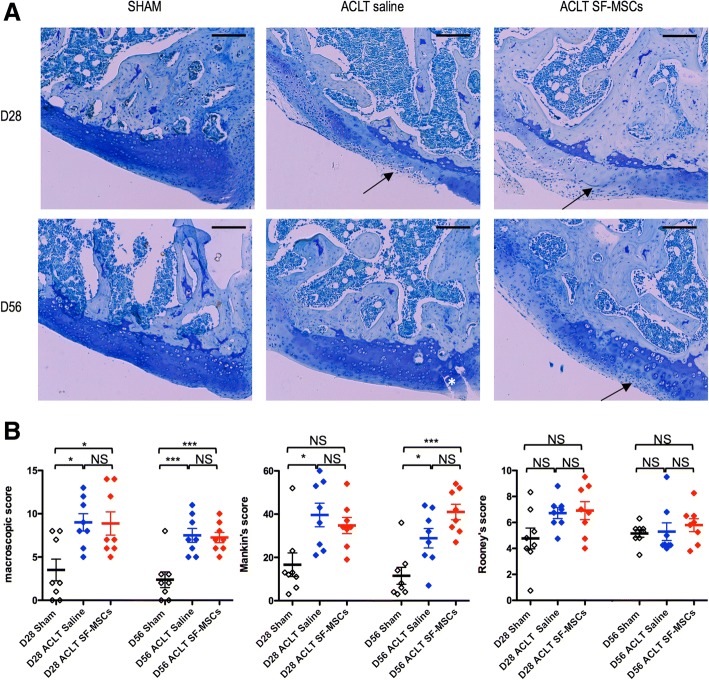
Methods: SF-MSCs were isolated from human SF aspirates in patients suffering from advanced OA undergoing total knee joint replacements. SF-MSCs at passage 2 (P2) were characterized by flow cytometry for epitope profiling. SF-MSCs at P2 were subsequently cultured in vitro to assess their multilineage potentials. To assess their chondrogenicity, SF-MSCs at P4 were seeded in collagen sponges for 4 weeks under various oxygen tensions and growth factors combinations to estimate their gene profile and matrix production. Also, SF-MSCs were injected into the joints in a nude rat anterior cruciate ligament transection (ACLT) to macroscopically and histologically assess their possible chondroprotective properties,.

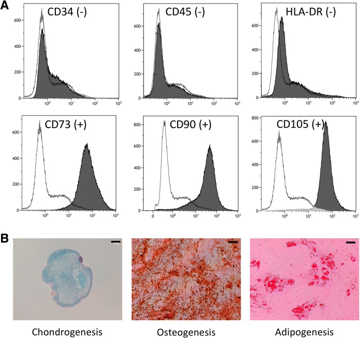
Results: We characterized the stemness (CD73+, CD90+, CD105+, CD34-, CD45-) and demonstrated the multilineage potency of SF-MSCs in vitro. Furthermore, the chondrogenic induction (TGF-ß1 ± BMP-2) of these SF-MSCs in collagen sponges demonstrated a good capacity of chondrogenic gene induction and extracellular matrix synthesis. Surprisingly, hypoxia did not enhance matrix synthesis, although it boosted chondrogenic gene expression (ACAN, SOX9, COL2A1). Besides, intra-articular injections of xenogenic SF-MSCs did exert neither chondroprotection nor inflammation in ACLT-induced OA in the rat knee.

Conclusions: Advanced OA SF-MSCs seem better candidates for cell-based constructs conceived for cartilage defects rather than intra-articular injections for diffuse OA.

Keywords: Cartilage; Collagen sponge; Stem cells; Synovial fluid; Tissue engineering.


Conflict of interest statement

Ethics approval and consent to participate

The clinical protocol was approved by the ethical committee of our university hospital. The experimental study in the rat was approved by our local Ethical Committee for the Animal studies and validated by the French Ministry of National Education, Higher Education and Research.

Consent for publication

Not applicable.

Competing interests

The authors declare that they have no competing interests.

Publisher’s Note

Springer Nature remains neutral with regard to jurisdictional claims in published maps and institutional affiliations.

Figures

Fig. 1
Fig. 1
Immunophenotype and trilineage differentiation of fibroblastic cells derived from advanced OA synovial fluid. a Surface marker expression of human MSCs derived from advanced OA synovial fluid at passage 2. Each histogram is a representative result of three SF-MSC samples. The white histogram represents negative control response, and black histogram is for the samples. Results showed negativity for CD34, CD45, and HLA-DR and positivity for MSCs markers CD90, CD105, and CD73. b Chondrogenic, osteogenic, and adipogenic potentials of SF-MSCs. The chondrogenic differentiation of SF-MSCs was assessed by alcian blue staining on pellet cultured during 28 days in chondrogenic medium (TGF-β1). Cells were cultured in osteogenic medium during 14 days, and calcium mineralization was evaluated by Alizarin Red S staining. Lipid droplets were observed in SF-MSCs cultured in adipogenic induction medium for 28 days with Red Oil staining. A representative example for two samples is shown. Scale bars 200 μm
Fig. 2
Fig. 2
Effect of culture conditions on the mRNA relative expression in cartilage engineered substitutes. Synovial fluid derived mesenchymal stem cells (sf-MSCs) were cultured in collagen sponges in normoxia (21% O2—gray histogram) or in hypoxia (5% O2—white histogram) during 28 days. Sponges were treated from day 3 with ITS 1% as control condition or with TGF-β1 (10 ng/mL) alone or in combination with BMP-2 (100 ng/mL) and BMP-2 (100 ng/mL) alone. We investigated relative mRNA expression by using real-time polymerase chain reaction of chondrogenic (COMP, ACAN, SOX9, COL2A1, COL2B), fibrotic (VCAN, COL1A1) and osteogenic markers (BGLAP and RUNX2). All results were normalized to RPS29 mRNA expression. Each histogram represents three independent experiments performed in triplicate. Data are presented as mean ± standard error of the mean. Statistical analysis was performed initially with a one-way ANOVA with a Dunnett’s post hoc test versus their internal control ITS for each condition (normoxia separated from hypoxia) followed by a two-way ANOVA with a Bonferroni’s post hoc test on all the values the interaction of hypoxia. Asterisks represent significant difference versus the control condition (ITS 1%) *p < 0.05; **p < 0.01; ***p < 0.001 in each condition. Hash sign represents significant difference of growth factor condition in normoxia versus hypoxia taken into account both conditions #p < 0.05; ##p < 0.01; ###p < 0.001 (significant interaction of hypoxia)
Fig. 3
Fig. 3
Histological, immunohistochemical analyses and GAG contents of cartilage engineered substitutes produced using human SF-MSCs and collagen sponge at D28 under various oxygen conditions and various growth factors. All observations were carried out on three different sponges for each culture condition and for each patient. The differences between three sponges of each group are very low; we choose to submit only one photograph. The sections were stained with alcian blue to reveal proteoglycan content (a) and immuno-histochemical analyses against type II collagen (Col2) was performed (c). The scale bars represent 400 μm. b and d represents densitometry measurement of alcian Blue and type II collagen respectively. Concentration of GAGs was measured inside sponges with a colorimetric assay using dimethylmethylene blue (e). Statistical analysis was performed initially with a one-way ANOVA with a Dunnett’s post hoc test versus their internal control ITS for each condition (normoxia separated from hypoxia) followed by a two-way ANOVA with a Bonferroni’s post hoc test on all the values the interaction of hyopoxia. Asterisks represent significative difference versus the control condition (ITS 1%) *p < 0.05; **p < 0.01; ***p < 0.001 (c, d, and e). There is no significant interaction of hypoxia
Fig. 4
Fig. 4
Histological and macroscopic scoring of experimental OA rat model induced by anterior cruciate ligament transection (ACLT) at days 28 and 56 after surgery. a Histological evaluation of medial femur by toluidine blue staining. Arrow represents surface irregularities or decrease of matrix contents, asterisk indicate fissure. Scale bars represent 200 μm. b represents macroscopic, Mankin’s, and Rooney’s scores of whole studied right knees. Individual values and mean ± SEM from eight rats per batch are represented (D28 and D56 SHAM in black; D28 and D56 ACLT + saline in blue; D28 and D56 SF-MSCs in red). Statistical analysis was performed by a one-way ANOVA with a Bonferroni’s post hoc test for multiple comparisons. Asterisks represent significant difference *p < 0.05; ***p < 0.001

Similar articles

Cited by

References

    1. Yasui Y, Ando W, Shimomura K, Koizumi K, Ryota C, Hamamoto S, et al. Scaffold-free, stem cell-based cartilage repair. J Clin Orthop Trauma. 2016;7(3):157–163. doi: 10.1016/j.jcot.2016.06.002. - DOI - PMC - PubMed
    1. Harris JD, Siston RA, Brophy RH, Lattermann C, Carey JL, Flanigan DC. Failures, re-operations, and complications after autologous chondrocyte implantation--a systematic review. Osteoarthr Cartil. 2011;19(7):779–791. doi: 10.1016/j.joca.2011.02.010. - DOI - PubMed
    1. Zingler C, Carl HD, Swoboda B, Krinner S, Hennig F, Gelse K. Limited evidence of chondrocyte outgrowth from adult human articular cartilage. Osteoarthr Cartil. 2016;24(1):124–128. doi: 10.1016/j.joca.2015.07.014. - DOI - PubMed
    1. Ma B, Leijten JC, Wu L, Kip M, van Blitterswijk CA, Post JN, et al. Gene expression profiling of dedifferentiated human articular chondrocytes in monolayer culture. Osteoarthr Cartil. 2013;21(4):599–603. doi: 10.1016/j.joca.2013.01.014. - DOI - PubMed
    1. Koyama N, Okubo Y, Nakao K, Osawa K, Fujimura K, Bessho K. Pluripotency of mesenchymal cells derived from synovial fluid in patients with temporomandibular joint disorder. Life Sci. 2011;89(19–20):741–747. doi: 10.1016/j.lfs.2011.09.005. - DOI - PubMed

Publication types