

Case Reports
doi: 10.30802/AALAS-CM-18-000004.
Epub 2018 Nov 28.
Longitudinal MRI Evaluation of Ischemic Stroke in the Basal Ganglia of a Rhesus Macaque (Macaca mulatta) with Seizures
Affiliations
- PMID: 30486918
- PMCID: PMC6310199
- DOI: 10.30802/AALAS-CM-18-000004
Item in Clipboard
Case Reports
Longitudinal MRI Evaluation of Ischemic Stroke in the Basal Ganglia of a Rhesus Macaque (Macaca mulatta) with Seizures
Comp Med.
.
Abstract
An adult rhesus macaque developed seizures after the induction of ischemic stroke. Initially, on the day of surgery, a focal ischemic lesion was present exclusively in the right caudate nucleus. By 48 h after stroke induction, the lesion had extended into the putamen, when a seizure was observed. Our report highlights the temporal changes in infarction of unilateral basal ganglia after acute stroke and the accompanying clinical symptoms. This unusual case may provide additional information regarding the involvement of the basal ganglia in seizures, given that prior case reports and studies usually have not described the temporal and spatial evolution of the lesion before clinical symptoms emerge.
Figures
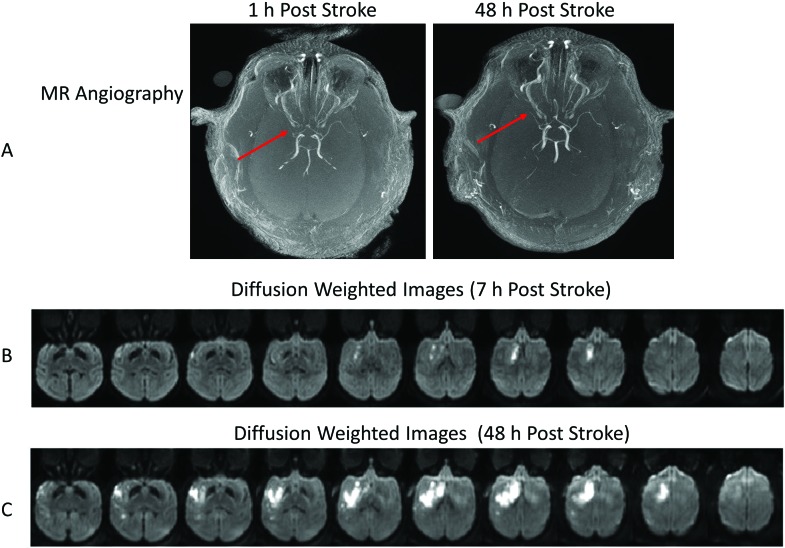
MR angiography and diffusion-weighted images of the rhesus macaque that developed seizures after stroke (animal ID RPF6). (A) Axial MR angiographic images at 1 and 48 h after stroke show the occluded MCA (arrows). Diffusion-weighted imaging highlights the evolution of the infarct at (B) 7 h and (C) 48 h after MCA occlusion.
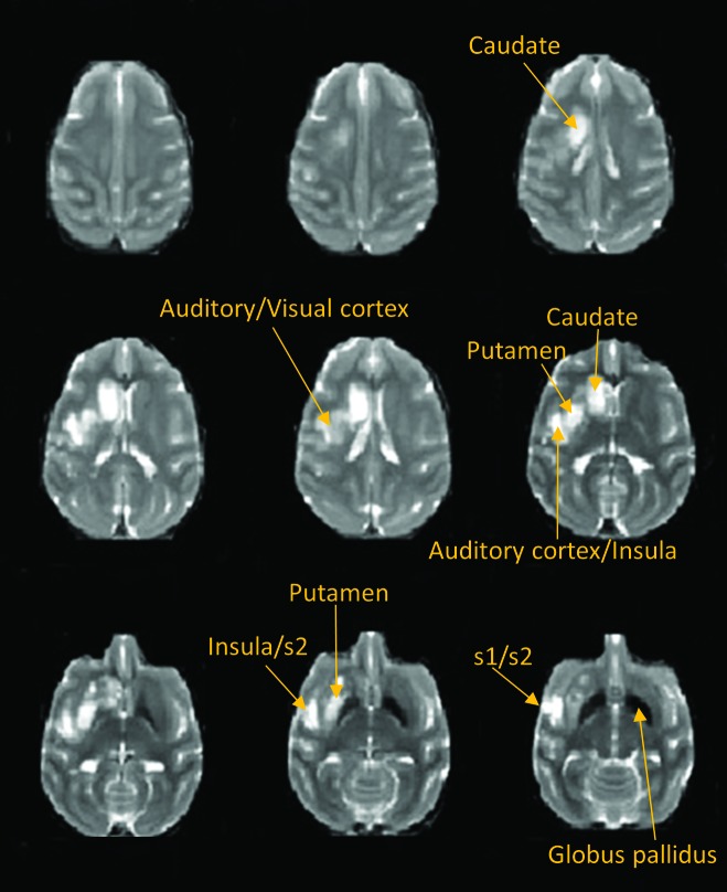
These T2-weighted images at 48 h after stroke illustrate the presence of edema after the stroke insult in our macaque.
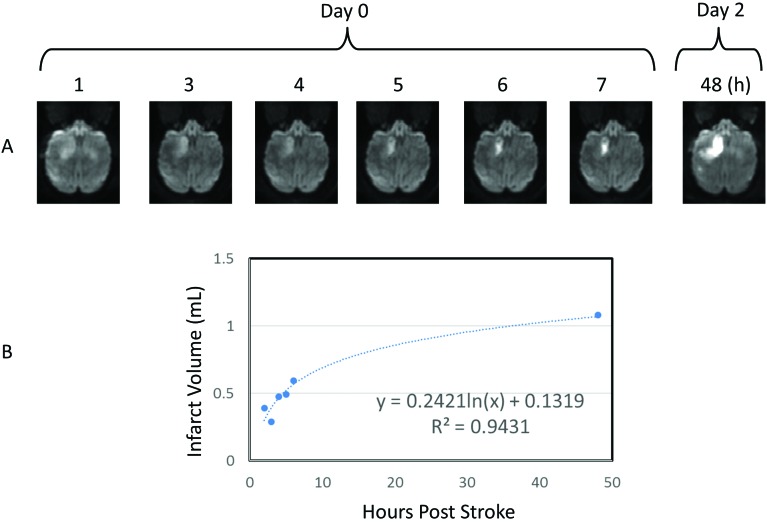
Temporal and spatial evolution of the stroke lesion in the basal ganglia. Single panels show the development of infarction from 1 to 48 h after surgery (upper panel). The progressive changes of entire infarction volumes fit a natural logarithmic pattern (bottom panel).
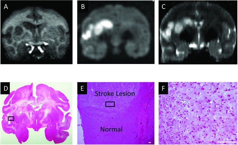
Demonstration of the stroke lesion in the macaque's brain by using MRI and neuropathology. (A) T1-weighted coronal image. (B) Diffusion-weighted coronal images. (C) T2-weighted coronal image. (D) Hematoxylin and eosin staining. (E) Magnification of the box in panel D. Scale bar, 100 µm. (F) Magnification of the box in panel E. Scale bar, 100 µm.
References
-
- Animal Welfare Act as Amended. 2013. 7 USC §2131–2159.
