. 2019 Jan 4;146(1):dev170225.
doi: 10.1242/dev.170225.

Two functionally redundant sources of retinoic acid secure spermatogonia differentiation in the seminiferous epithelium

Affiliations

Two functionally redundant sources of retinoic acid secure spermatogonia differentiation in the seminiferous epithelium

Marius Teletin et al. Development. .

Abstract

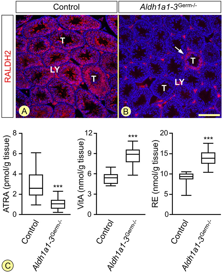
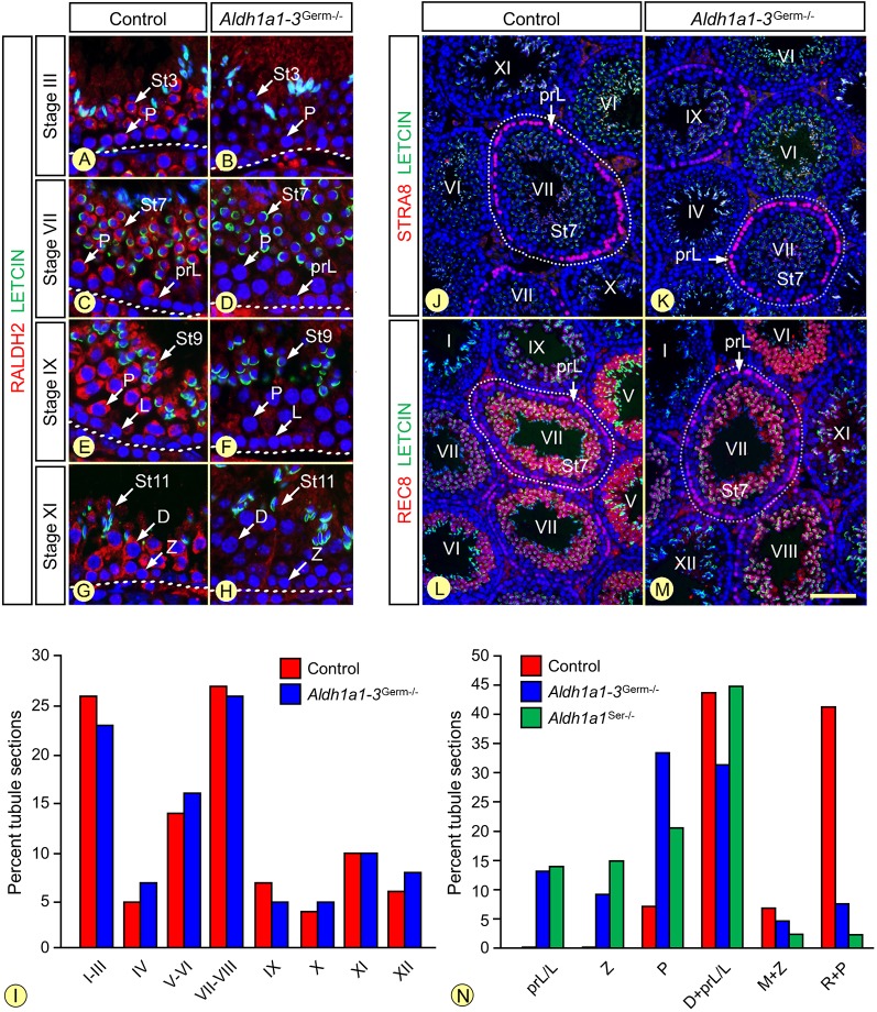
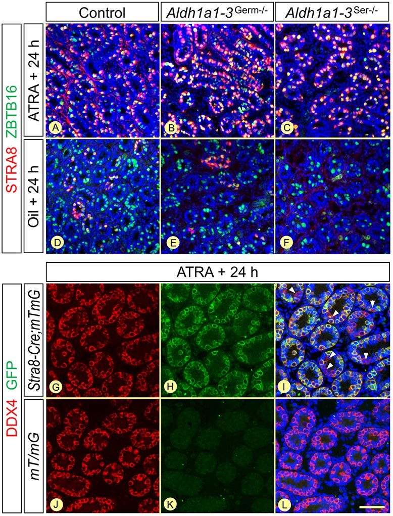
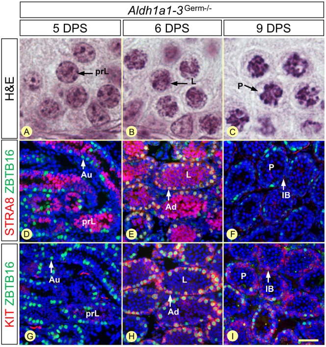
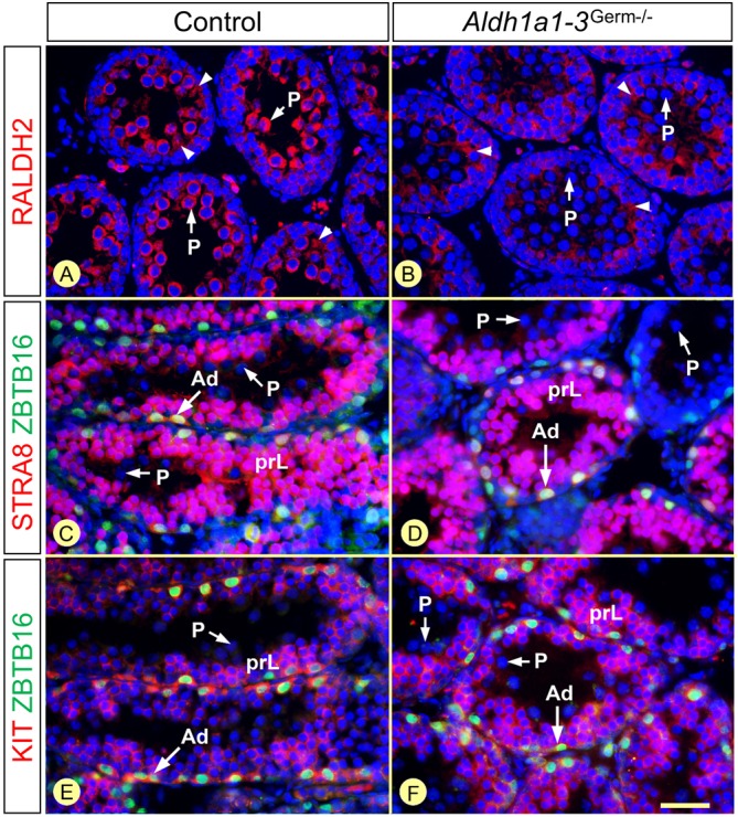
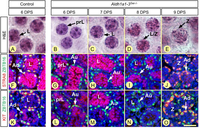
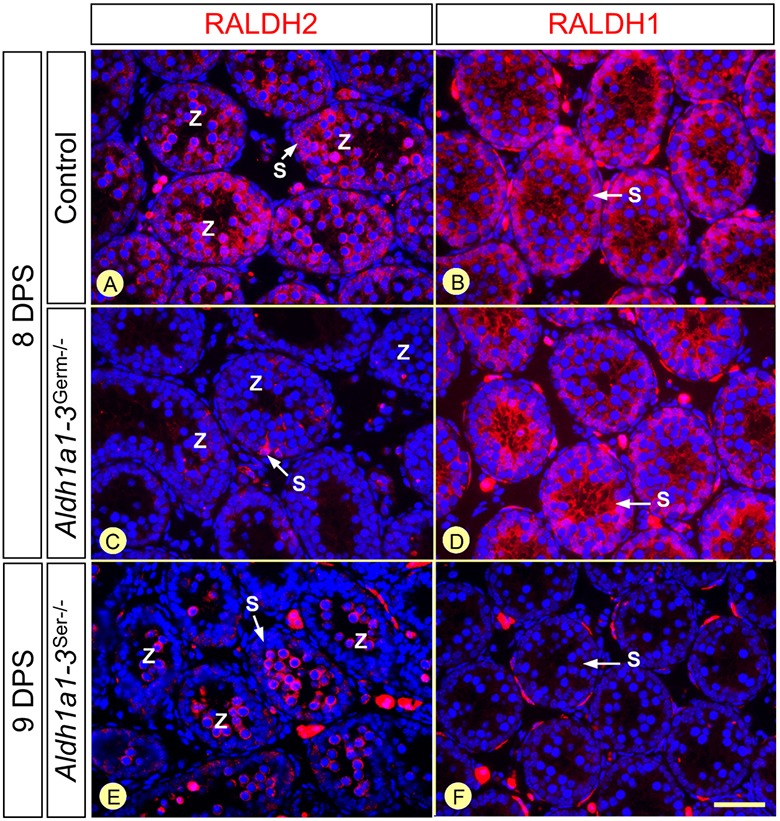
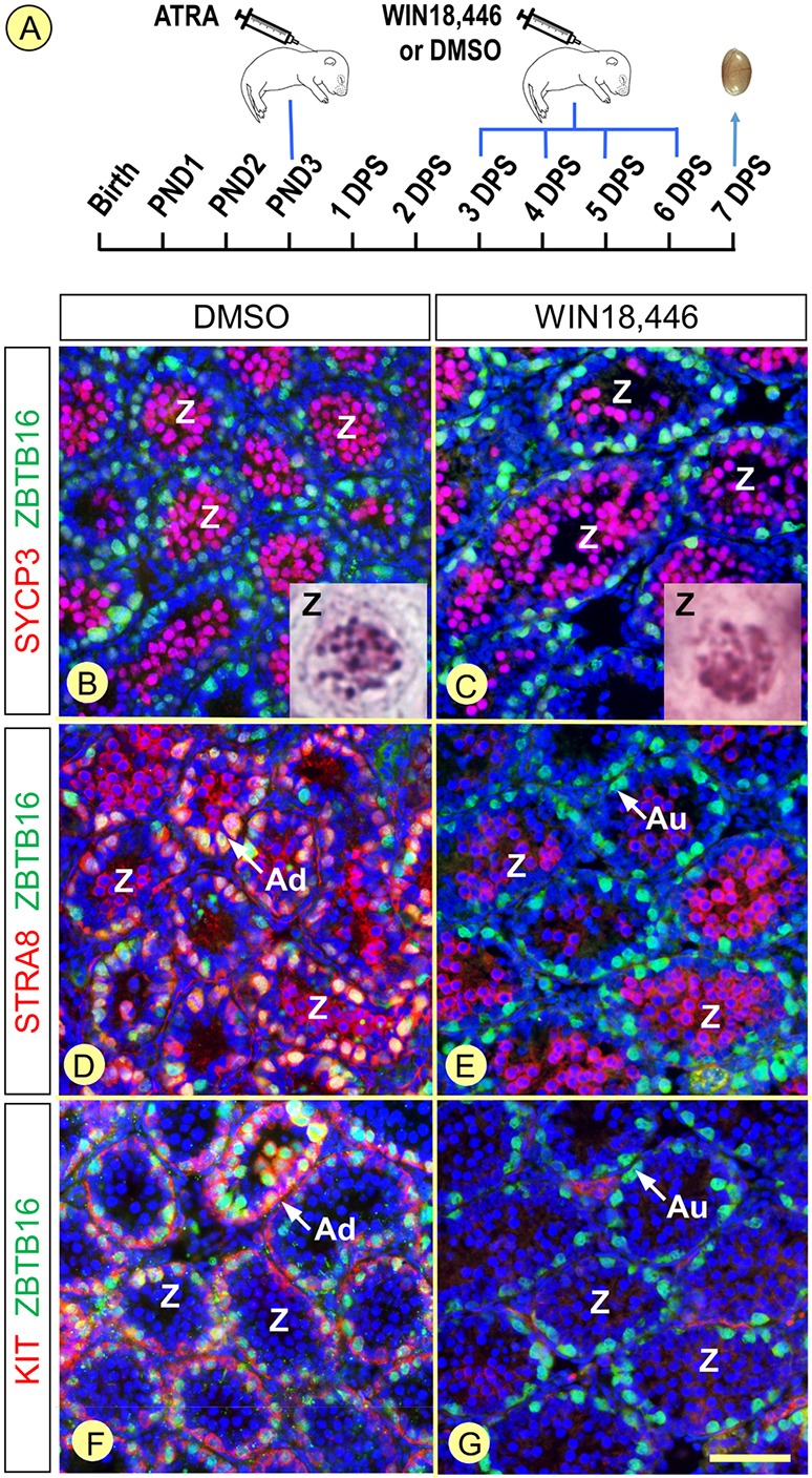
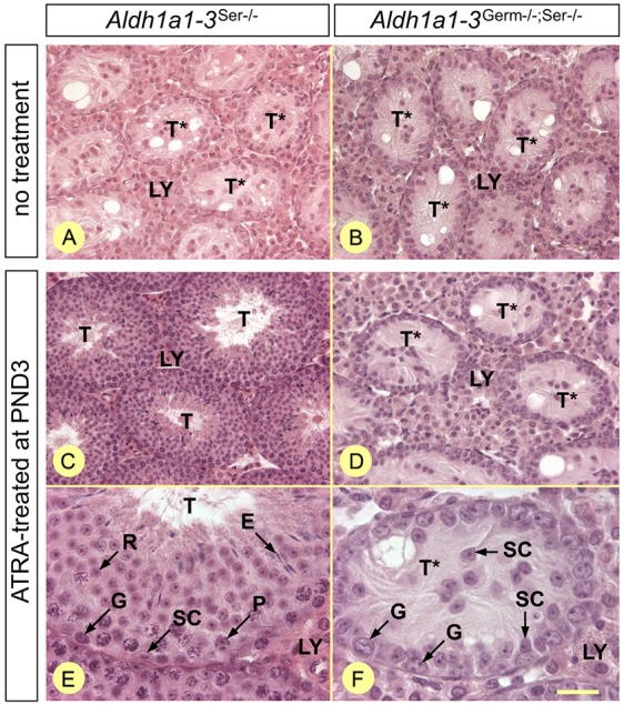
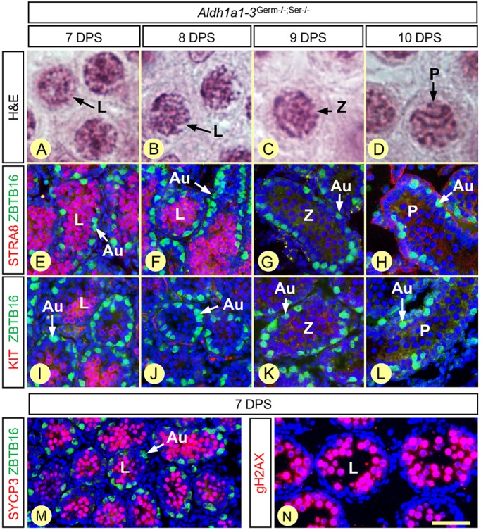
In mammals, all-trans retinoic acid (ATRA) is instrumental to spermatogenesis. It is synthesized by two retinaldehyde dehydrogenases (RALDH) present in both Sertoli cells (SCs) and germ cells (GCs). In order to determine the relative contributions of each source of ATRA, we have generated mice lacking all RALDH activities in the seminiferous epithelium (SE). We show that both the SC- and GC-derived sources of ATRA cooperate to initiate and propagate spermatogenetic waves at puberty. In adults, they exert redundant functions and, against all expectations, the GC-derived source does not perform any specific roles despite contributing to two-thirds of the total amount of ATRA present in the testis. The production from SCs is sufficient to maintain the periodic expression of genes in SCs, as well and the cycle and wave of the SE, which account for the steady production of spermatozoa. The production from SCs is also specifically required for spermiation. Importantly, our study shows that spermatogonia differentiation depends upon the ATRA synthesized by RALDH inside the SE, whereas initiation of meiosis and expression of STRA8 by spermatocytes can occur without ATRA.

Keywords: Aldh8a1 knockout; Immunohistochemistry; Lgals1; Puberty; Spermatogenesis.


Conflict of interest statement

Competing interestsThe authors declare no competing or financial interests.

Figures

Fig. 1.
Fig. 1.
The Aldh1a1-3Germ−/− mutation suppresses RALDH2 expression in germ cells and alters testicular retinoid metabolism. (A,B) Immunohistochemical detection of RALDH2 (red signal) on histological sections from 8-week-old control and Aldh1a1-3Germ−/− mice. The white arrow in B indicates the only seminiferous tubule (T) cross-section, out of the 230, that still expresses RALDH2. The signal observed in Leydig cells (LY) is unspecific, as RALDH2 transcripts are not detected in these cells (Vernet et al., 2006b). Nuclei were counterstained with DAPI (blue signal). Scale bar: 160 µm. (C) Box-and-whisker plots showing the testicular concentrations of ATRA, vitA and retinyl esters (RE) in 9- to 10-week-old Aldh1a1-3Germ−/− (n=16) and control (n=14) mice. The boxes indicate the upper and lower quartiles, the lines inside mark the medians, and the whiskers delineate the highest and lowest values. ***P<0.001, Student's t-test.
Fig. 2.
Fig. 2.
The adult Aldh1a1-3Germ−/− mutant SE is normal. (A-H) Detection of RALDH2 (red signal) on histological sections from 8-week-old control and Aldh1a1-3Germ−/− mice. RALDH2 is completely absent in the mutant GC. (I) Relative frequencies of the stages of the SE in 9- to 10-week-old control (red bars, n=10) and Aldh1a1-3Germ−/− (blue bars, n=4) testes. (J-M) Detection of STRA8 and REC8 (red signals) on histological sections from 8-week-old control and Aldh1a1-3Germ−/− mice. In both cases, STRA8 is expressed in preleptotene and leptotene spermatocytes at epithelial stages VII to IX. REC8 is detected in preleptotene spermatocytes at stages VII and VIII, and in spermatids at stages V to IX. Nuclei were counterstained by DAPI (blue signal) and the acrosomal system by Alexa Fluor 488-conjugated peanut agglutinin (green signal), allowing proper staging. Roman numerals designate stages of the SE cycle. The dotted lines indicate the periphery of seminiferous tubules. (N) Ablation of either the SC- or the GC-derived source of ATRA delays spermatogenesis. Mean percentages of seminiferous tubule sections in P21 control (red bars), Aldh1a1-3Germ−/− (blue bars) and Aldh1a1Ser−/− (green bars) testes containing: preleptotene or leptotene spermatocytes and a peripheral row of spermatogonia (prL/L); zygotene spermatocytes and a peripheral row of spermatogonia (Z); early pachytene spermatocytes and a peripheral row of spermatogonia (P); late pachytene or diplotene spermatocytes with a peripheral row of either preleptotene or leptotene spermatocytes (D+prL/L); meiotic metaphases with a peripheral row of zygotene spermatocytes (M+Z); round spermatids with a peripheral row of pachytene spermatocytes (R+P). D, prL, L, P and Z indicate diplotene, preleptotene, leptotene, pachytene and zygotene spermatocytes, respectively; St3 to St11, steps of spermatid maturation. Scale bar: 30 µm in A-H; 80 µm in J-M.
Fig. 3.
Fig. 3.
Synchronization of differentiation and Cre-mediated excision in spermatogonia. (A-F) Detection of STRA8 (red signal) and ZBTB16 (green nuclear signal) on histological sections from control, Aldh1a1-3Germ−/− and Aldh1a1-3Ser−/− testes 24 h after the administration of ATRA or sunflower oil at P3. (G-L) Detection of DDX4 (red cytoplasmic signal) and GFP (green membranous signal) on histological sections from Stra8-Cre;mT/mG and mT/mG mice 24 h after the administration of ATRA at P3. Efficient mT/mG excision by Stra8-Cre is assessed by GFP expression in almost all spermatogonia (G-I). In contrast, GFP is never detected in the absence of Stra8-Cre (J-L). The arrowheads in I indicate a few spermatogonia in which mTmG is not excised. Scale bar: 80 µm in A-F; 70 µm in G-L.
Fig. 4.
Fig. 4.
Chronology of GC differentiation in Aldh1a1-3Germ−/− testes 5, 6 and 9 days after synchronization. (A-C) Morphology of the spermatocytes, present at the center of the seminiferous tubules, assessed by Hematoxylin and Eosin staining. (D-I) Characterization of the spermatogonia populations, present at the periphery of the seminiferous tubules, by detection of ZBTB16 (green signal) and either STRA8 or KIT (red signals), as indicated. Nuclei were counterstained with DAPI (blue signal). D and G, and E and H are adjacent sections. Au, Ad and IB indicate Aundiff, Adiff and intermediate or B spermatogonia, respectively; DPS, day post-synchronization; prL, L and P indicate preleptotene, leptotene and pachytene spermatocytes, respectively. Scale bar: 6 µm in A-C; 60 µm in D-I.
Fig. 5.
Fig. 5.
The same types of GC are present in control and Aldh1a1-3Germ−/− testes 12 days after synchronization. (A,B) Detection of RALDH2 (red signal). (C-F) Characterization of the spermatogonia and spermatocyte populations, present at the periphery of the seminiferous tubules, by detection of ZBTB16 (green signal) and either STRA8 or KIT (red signals), as indicated. Nuclei were counterstained with DAPI (blue signal). C and E are consecutive sections. Ad indicates Adiff spermatogonia; prL and P indicate preleptotene and pachytene spermatocytes, respectively. Arrowheads indicate RALDH2 in the cytoplasm of SCs. Scale bar: 30 µm.
Fig. 6.
Fig. 6.
Chronology of GC differentiation in Aldh1a1-3Ser−/− testes at 6, 7, 8 and 9 days after synchronization. (A-E) Morphology of the spermatocytes, present at the center of the seminiferous tubules, assessed by Hematoxylin and Eosin staining in control (A) and mutant (B-E) testes. (F-O) Characterization of the spermatogonia populations, present at the periphery of the seminiferous tubules, assessed by detection of ZBTB16 (green signal) and either STRA8 or KIT (red signals), as indicated, in control (F,K) and mutant (G-J, L-O) testes. Nuclei were counterstained with DAPI (blue signal). Au and Ad indicate Aundiff and Adiff spermatogonia, respectively; DPS, day post-synchronization; prL, L and Z indicate preleptotene, leptotene and zygotene spermatocytes, respectively. Scale bar: 6 µm in A-E; 60 µm in F-O.
Fig. 7.
Fig. 7.
Zygotene spermatocytes are the first GC type to express RALDH2 during testis development. (A-F) Detection of RALDH1 and RALDH2 (red signals) at 8 or 9 DPS in Aldh1a1-3Germ−/−, Aldh1a1-3Ser−/− and control testes, as indicated. Nuclei were counterstained with DAPI (blue signal). Z, zygotene spermatocytes; S, Sertoli cell cytoplasm. Scale bar: 50 µm.
Fig. 8.
Fig. 8.
Inhibition of spermatogonia differentiation, but not meiosis, upon exposure of synchronized Aldh1a1-3Germ−/− testes to WIN18,446. (A) Pups were treated with ATRA at P3, with WIN18,446 or DMSO between 3 and 6 DPS, and their testes were analyzed at 7 DPS. (B,C) Spermatocytes expressing SYCP3 (red signal) and showing nuclear morphologies corresponding to zygotene spermatocytes (insets) fill the seminiferous tubules in both DMSO- and WIN18,446-treated pups. (D,E) STRA8-positive spermatogonia are present in DMSO-treated but totally absent in WIN18,446-treated pups (red signal at the periphery of the tubules). In contrast, STRA8 is expressed in zygotene spermatocytes in both DMSO- and WIN18,446-treated pups (red signal at the center of the tubules). (F,G) KIT-positive spermatogonia (red signal) are present in DMSO-treated but totally absent in WIN18,446-treated pups. Spermatogonia nuclei are stained using ZBTB16 (green signals), while all cell nuclei are stained using DAPI (blue signals). Au and Ad indicate Aundiff and Adiff spermatogonia, respectively. Z, zygotene spermatocytes. Scale bar: 60 µm.
Fig. 9.
Fig. 9.
Neonatal exposure of Aldh1a1-3Germ−/−;Ser−/− mutants to exogenous ATRA cannot sustain spermatogenesis. (A,B) Histological sections of testes from untreated 6-week-old mutant males. (C-F) Histological sections 6 weeks after a single injection of ATRA at P3. Genotypes are as indicated. All sections were stained with Hematoxylin and Eosin. E, elongated spermatids; G, spermatogonia; LY, Leydig cells; P, pachytene spermatocytes; R, round spermatids; SC, Sertoli cells; T and T*, tubule cross-sections displaying spermatogenesis or only Sertoli cells and spermatogonia, respectively. Scale bar: 60 µm in A-D; 20 µm in E,F.
Fig. 10.
Fig. 10.
Chronology of GC differentiation in Aldh1a1-3Germ−/−;Ser−/− testes at 7, 8, 9 and 10 days after synchronization. (A-D) Morphology of the spermatocytes, present at the center of the seminiferous tubules, assessed by Hematoxylin and Eosin staining. (E-L) Characterization of the spermatogonia populations, present at the periphery of the seminiferous tubules, assessed by detection of ZBTB16 (green signal) and either STRA8 or KIT (red signals), as indicated. (M,N) Expression of the meiotic markers SYCP3 and gH2AX (red signals) in leptotene spermatocytes. Nuclei were counterstained with DAPI (blue signal). Au, Aundiff spermatogonia; DPS, day post-synchronization; L, Z and P indicate leptotene zygotene and pachytene spermatocytes, respectively. Scale bar: 6 µm in A-D; 60 µm in E-L; 80 µm in M; 40 µm in N.

References

    1. Anderson E. L., Baltus A. E., Roepers-Gajadien H. L., Hassold T. J., de Rooij D. G., van Pelt A. M. M. and Page D. C. (2008). Stra8 and its inducer, retinoic acid, regulate meiotic initiation in both spermatogenesis and oogenesis in mice. Proc. Natl. Acad. Sci. USA 105, 14976-14980. 10.1073/pnas.0807297105 - DOI - PMC - PubMed
    1. Arnold S. L. M., Kent T., Hogarth C. A., Griswold M. D., Amory J. K. and Isoherranen N. (2015). Pharmacological inhibition of ALDH1A in mice decreases all-trans retinoic acid concentrations in a tissue specific manner. Biochem. Pharmacol. 95, 177-192. 10.1016/j.bcp.2015.03.001 - DOI - PMC - PubMed
    1. Beedle M.-T., Stevison F., Zhong G., Topping T., Hogarth C., Isoherranen N. and Griswold M. D. (2018). Sources of all-trans retinal oxidation independent of the aldehyde dehydrogenase 1A isozymes exist in the postnatal testis. Biol. Reprod. 10.1093/biolre/ioy200 - DOI - PMC - PubMed
    1. Blume-Jensen P., Jiang G., Hyman R., Lee K.-F., O'Gorman S. and Hunter T. (2000). Kit/stem cell factor receptor-induced activation of phosphatidylinositol 3′-kinase is essential for male fertility. Nat. Genet. 24, 157-162. 10.1038/72814 - DOI - PubMed
    1. Bouillet P., Sapin V., Chazaud C., Messaddeq N., Décimo D., Dollé P. and Chambon P. (1997). Developmental expression pattern of Stra6, a retinoic acid-responsive gene encoding a new type of membrane protein. Mech. Dev. 63, 173-186. 10.1016/S0925-4773(97)00039-7 - DOI - PubMed

Publication types