. 2018 Dec 11;115(50):E11642-E11650.
doi: 10.1073/pnas.1808731115. Epub 2018 Nov 28.

UBL domain of Usp14 and other proteins stimulates proteasome activities and protein degradation in cells

Affiliations

UBL domain of Usp14 and other proteins stimulates proteasome activities and protein degradation in cells

Hyoung Tae Kim et al. Proc Natl Acad Sci U S A. .

Erratum in

Abstract

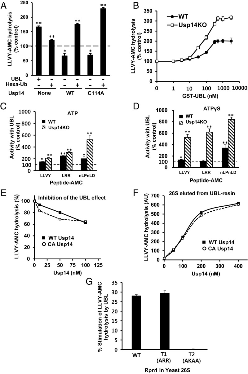
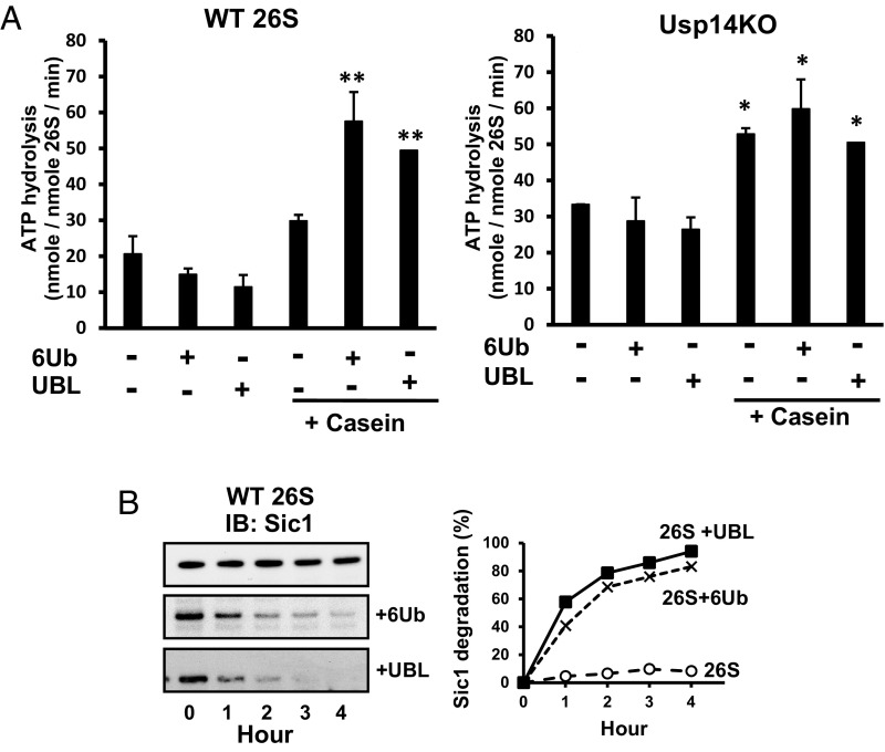
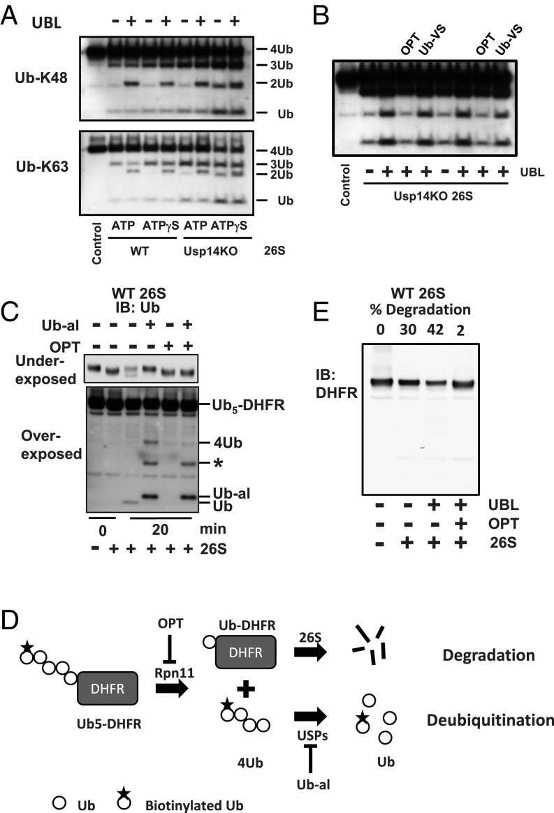
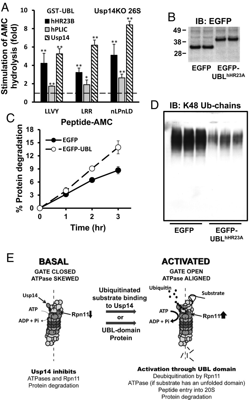
The best-known function of ubiquitin-like (UBL) domains in proteins is to enable their binding to 26S proteasomes. The proteasome-associated deubiquitinating enzyme Usp14/UBP6 contains an N-terminal UBL domain and is an important regulator of proteasomal activity. Usp14 by itself represses multiple proteasomal activities but, upon binding a ubiquitin chain, Usp14 stimulates these activities and promotes ubiquitin-conjugate degradation. Here, we demonstrate that Usp14's UBL domain alone mimics this activation of proteasomes by ubiquitin chains. Addition of this UBL domain to purified 26S proteasomes stimulated the same activities inhibited by Usp14: peptide entry and hydrolysis, protein-dependent ATP hydrolysis, deubiquitination by Rpn11, and the degradation of ubiquitinated and nonubiquitinated proteins. Thus, the binding of Usp14's UBL (apparently to Rpn1's T2 site) seems to mediate the activation of proteasomes by ubiquitinated substrates. However, the stimulation of these various activities was greater in proteasomes lacking Usp14 than in wild-type particles and thus is a general response that does not involve some displacement of Usp14. Furthermore, the UBL domains from hHR23 and hPLIC1/UBQLN1 also stimulated peptide hydrolysis, and the expression of hHR23A's UBL domain in HeLa cells stimulated overall protein degradation. Therefore, many UBL-containing proteins that bind to proteasomes may also enhance allosterically its proteolytic activity.

Keywords: UBL domain; Usp14/Ubp6; hHR23/Rad23; hPLIC/ubiquilin; proteasome activation.


Conflict of interest statement

The authors declare no conflict of interest.

Figures

Fig. 1.
Fig. 1.
By binding to 26S proteasomes, Usp14’s UBL domain stimulates peptide cleavage by all three peptidase sites, especially in proteasomes lacking Usp14. Activities of WT and Usp14KO 26S were assayed by measuring cleavage of the fluorogenic peptide-amc substrates (10 μM) in the presence of ATP or ATPγS (100 μM). Control (100%) indicates the peptidase activity of each type of proteasome (1 nM) without added proteins. (A) Addition of GST-UBL from Usp14 (200 nM) by itself stimulates the chymotrypsin-like activity of 26S similar to addition of Usp14 (1 μM) plus a linear hexa-Ub chain (200 nM). Addition of WT Usp14 or the enzymatically inactive C114A mutant by themselves inhibited, but together with hexa-Ub stimulated, peptidase activity, especially with the inactive Usp14 mutant. LLVY-amc: Suc-Leu-Leu-Val-Tyr-amc as the substrate for the chymotrypsin-like activity. *P < 0.05 and **P < 0.01 by Student’s t test. Data are the means ± SD; n = 4. (B) The UBL of Usp14 stimulates the chymotrypsin-like activity of both WT and Usp14KO 26S. The UBL seems to bind with similar affinities (∼100 nM) to both, but causes a much larger stimulation of the Usp14KO 26S. Data are the means ± SD; n = 5. (C) All three peptidase activities are stimulated more by the UBL (200 nM) in the Usp14KO 26S than in the WT with ATP present. LRR-amc: Boc-Leu-Arg-Arg-amc as the substrate for the trypsin-like activity; nLPnLD-amc: Ac-Nle-Pro-Nle-Asp-amc as the substrate for the caspase-like activity. *P < 0.05 and **P < 0.01 by Student’s t test. Data are the means ± SD; n = 6. (D) In the presence of ATPγS, the UBL domain further enhanced (five- to eightfold) the three peptidase activities of Usp14KO 26S. Peptide hydrolysis by WT 26S was also stimulated, but to a smaller extent. *P < 0.05 and **P < 0.01 by Student’s t test. Data are the means ± SD; n = 6. (E) Full-length Usp14 inhibits the activation of peptide hydrolysis by UBL. Addition of increasing concentrations of WT Usp14 or the inactive C114A mutant decreased the chymotrypsin-like activity of 26S proteasomes in the presence of UBL and ATPγS. Usp14KO proteasomes (1 nM) were preincubated with the UBL (100 nM), and different concentrations of Usp14 were then added to the mixture. Data are the means ± SD; n = 4. (F) Full-length Usp14 competes with the GST-UBL for binding to the 26S proteasome. Usp14KO 26S bound to UBL on resin was eluted by adding increasing concentrations of Usp14 (55). Proteasomes eluted were quantitated using LLVY-amc hydrolysis (20). Data are represented as means ± SD; n = 6. (G) The UBL domain from Usp14 stimulates the chymotrypsin-like activity of yeast WT and T1 mutant (ARR) 26S but not of the T2 mutant (AKAA) (32). Activities of the resin-bound yeast 26S were measured after adding the UBL derived from mammalian Usp14 (200 nM). Data are the means ± SD; n = 3.
Fig. 2.
Fig. 2.
UBL enhances ATP hydrolysis by WT proteasomes provided an unstructured protein (casein) is also present and stimulates the degradation of nonubiquitinated proteins. (A) Upon binding of casein (1 μM) and a hexa-Ub chain (1 μM), which together mimic the binding of Ub conjugates (20), ATP hydrolysis by WT and Usp14KO 26S (20 nM) increased about two- to threefold. The UBL (500 nM) plus casein also increased ATP hydrolysis in both to a similar extent as hexa-Ub plus casein. However, UBL or hexa-Ub alone did not increase ATP hydrolysis by either type of proteasome. However, in the Usp14KO 26S, in the absence of UBL or hexa-Ub chain, casein alone could increase ATP hydrolysis. ATP hydrolysis was measured using the malachite green assay (62). 6Ub: linear hexa-Ub. *P < 0.05 and **P < 0.01 by Student’s t test. Data are represented as means ± SD; n = 3. (B) The UBL domain, like a Ub chain, stimulates the degradation of an intrinsically unstructured protein (Sic1) by 26S proteasomes. Effects of UBL (500 nM) or linear hexa-Ub chain (1 μM) on the degradation of PY-Sic1 (100 nM) by WT 26S (2 nM) were assayed by Western blotting. Proteasomes were incubated with the UBL or Ub chain at room temperature for 15 min before the reaction started. (B, Left) At the indicated times, the remaining Sic1 was measured. IB, immunoblotting. (B, Right) Degradation of Sic1 was measured using ImageJ software and plotted. Similar data were obtained in three independent experiments.
Fig. 3.
Fig. 3.
UBL domain stimulates both Ub-chain disassembly by Rpn11 and the degradation of Ub5-DHFR. (A) UBL increases the breakdown of K48 and K63 tetra-Ub chains (368 nM) by MEF 26S with ATP or ATPγS present. 26S proteasomes (5 nM) were incubated with UBL as indicated at room temperature for 15 min before the reaction, which was carried out at 37 °C for 20 min. The generation of tri, di-, and mono-Ubs was analyzed by Western blotting as previously reported (25). Similar data were obtained in three independent experiments. (B) The UBL stimulates breakdown of K48 tetra-Ub by WT 26S with ATP present, and this stimulation must involve Rpn11 because it was inhibited by o-phenanthroline (OPT; 1 mM) but not by Ub-vinyl sulfone (Ub-VS; 200 nM). Similar data were obtained in two independent experiments. (C) 26S proteasomes disassemble the Ub chain on ubiquitinated DHFR to generate monomeric Ubs. Upon inhibition of Usp14 and Uch37 by Ub-aldehyde (300 nM), tetra-Ub chains are disassembled by Rpn11, and the Rpn11 inhibitor OPT (1 mM) blocked completely deubiquitination of DHFR. Degradation of Ub5-DHFR (100 nM) by 26S proteasomes (2 nM) in 20 min was assayed by Western blotting. The asterisk indicates nonspecific signal from Ub-aldehyde. (D) Illustration of why degradation of Ub5-DHFR is dependent on Rpn11. Neither Usp14 nor Uch37 can catalyze disassembly of the tetra-Ub chain. Only after Rpn11 cleaves tetraubiquitin chains en bloc off the protein can these other deubiquitinating enzymes disassemble the chain. Therefore, degradation of Ub5-DHFR depends on Rpn11 activity. (E) Although WT 26S degrades Ub5-DHFR rapidly, the addition of UBL (500 nM) enhances this process further. However, inhibition of Rpn11 activity by OPT (1 mM) completely blocks degradation. Similar data were obtained in three independent experiments.
Fig. 4.
Fig. 4.
UBL domains of other UBL-UBA proteins, hHR23 and hPLIC1, also stimulate peptide hydrolysis by 26S proteasomes, and expression of EGFP-UBL increases the degradation of long-lived proteins in HeLa cells. (A) With ATPγS (100 μM) and Usp14KO 26S (1 nM), the UBL domain (500 nM) of hHR23B stimulates proteasomes’ three peptidase activities three- to fivefold, while the UBL domain of hPLIC1 stimulates them twofold or less. Peptidase activities were measured as in Fig. 1. *P < 0.05 and **P < 0.01 by Student’s t test. Data are the means ± SD; n = 6. (B) EGFP and EGFP-UBL derived from hHR23A were expressed in HeLa cells (48 h after transfection). (C) HeLa cells expressing EGFP-UBL degrade long-lived proteins faster than control cells. Proteins were labeled with [3H]phenylalanine (5 μCi/mL) for 24 h. After 1 h in chase medium (DMEM containing 2 mg/mL nonradioactive phenylalanine) to allow short-lived proteins to be degraded, the breakdown of cellular proteins was measured in fresh chase medium for up to 3 h. Data are the means ± SD; n = 3. (D) The content of ubiquitin conjugates in HeLa cell lysates was lower after expression of EGFP-UBL than after EGFP. Forty-eight hours after transfection, equal amounts of proteins (2 μg) in lysates were electrophoresed and then Western blotted using anti-Ub antibody. Similar results were obtained from two independent experiments. (E) Summary of Usp14’s allosteric regulation of proteasomal degradation of ubiquitinated proteins. Without a Ub-conjugate bound, Usp14 (through its USP domain) inhibits ATP hydrolysis, substrate entry into the 20S, and deubiquitination by Rpn11 (25). However, upon binding Ub chains, Usp14 undergoes major structural transitions (2), and its UBL domain activates peptide entry into 20S, Rpn11, and ATP hydrolysis, if an unfolded protein also binds. These actions promote efficient degradation of ubiquitinated substrates. This activated state should be maintained as long as Usp14 binds the Ub chain. These processes return to the quiescent basal state when the substrate is degraded or is deubiquitinated and dissociates. These allosteric actions are not dependent on Usp14’s catalytic activity, which functions to degrade the Ub chain and thus limits the duration of the active state (25). Also shown is the proposed activation of proteasomes by UBL-containing proteins, which should be independent of the presence of Usp14.

Similar articles

Cited by

References

    1. Rock KL, Farfán-Arribas DJ, Colbert JD, Goldberg AL. Re-examining class-I presentation and the DRiP hypothesis. Trends Immunol. 2014;35:144–152. - PMC - PubMed
    1. Collins GA, Goldberg AL. The logic of the 26S proteasome. Cell. 2017;169:792–806. - PMC - PubMed
    1. Kisselev AF, et al. The caspase-like sites of proteasomes, their substrate specificity, new inhibitors and substrates, and allosteric interactions with the trypsin-like sites. J Biol Chem. 2003;278:35869–35877. - PubMed
    1. Smith DM, et al. Docking of the proteasomal ATPases’ carboxyl termini in the 20S proteasome’s alpha ring opens the gate for substrate entry. Mol Cell. 2007;27:731–744. - PMC - PubMed
    1. Lander GC, Martin A, Nogales E. The proteasome under the microscope: The regulatory particle in focus. Curr Opin Struct Biol. 2013;23:243–251. - PMC - PubMed

Publication types

LinkOut - more resources