. 2018 Dec 11;115(50):E11807-E11816.
doi: 10.1073/pnas.1808064115. Epub 2018 Nov 28.

The adult oligodendrocyte can participate in remyelination

Affiliations

The adult oligodendrocyte can participate in remyelination

Ian D Duncan et al. Proc Natl Acad Sci U S A. .

Abstract

Endogenous remyelination of the CNS can be robust and restore function, yet in multiple sclerosis it becomes less complete with time. Promoting remyelination is a major therapeutic goal, both to restore function and to protect axons from degeneration. Remyelination is thought to depend on oligodendrocyte progenitor cells, giving rise to nascent remyelinating oligodendrocytes. Surviving, mature oligodendrocytes are largely regarded as being uninvolved. We have examined this question using two large animal models. In the first model, there is extensive demyelination and remyelination of the CNS, yet oligodendrocytes survive, and in recovered animals there is a mix of remyelinated axons interspersed between mature, thick myelin sheaths. Using 2D and 3D light and electron microscopy, we show that many oligodendrocytes are connected to mature and remyelinated myelin sheaths, which we conclude are cells that have reextended processes to contact demyelinated axons while maintaining mature myelin internodes. In the second model in vitamin B12-deficient nonhuman primates, we demonstrate that surviving mature oligodendrocytes extend processes and ensheath demyelinated axons. These data indicate that mature oligodendrocytes can participate in remyelination.

Keywords: adult oligodendrocyte; large animal models; remyelination.


Conflict of interest statement

The authors declare no conflict of interest.

Figures
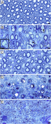
Fig. 1.
Fig. 1.
Mature myelin sheaths are seen in ventral columns at all stages of the disease and are mixed with demyelinated and remyelinated axons while the dorsal column initially has large areas of demyelination. (A) Normal mature white matter in the feline spinal cord ventral column. There is a mix of large, medium, and small diameter axons with appropriately thick myelin sheaths. (B) At the onset of disease in the ventral column, many axons become vacuolated, yet in each the axon remains intact (arrows). As myelin breaks down, intact axons can still be seen (arrowhead and high power shown in Inset). At this stage, occasional demyelinated and remyelinated axons are present. (C) In the ventral column of the recovered animal, there is a mix of remyelinated and mature myelin sheaths around axons of all diameters. (D) In the subpial area of the dorsal column of the same animal in B, there is complete demyelination with myelin debris. The axons are much smaller than in B as they are part of the fasciculus gracilis. (E) In the dorsal column of the same animal shown in C, all of the axons have thin remyelinated sheaths, different from the mosaic shown in C. (Scale bars, 20 µm.)
Fig. 2.
Fig. 2.
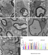
Oligodendrocytes have cytoplasmic connections to axons of variable diameter with thin or thick myelin sheaths. These five oligodendrocytes have cytoplasmic connections, seen on high power, contacting axons that have mature sheaths (g-ratios < 0.75) and thin myelin (remyelinated) sheaths (g-ratio > 0.75). Remyelinated myelin sheaths were seen around small diameter (AC and E) or large diameter (D) axons. (F) Definitive cytoplasmic connection is seen in an oligodendrocyte that myelinates a mature myelin sheath (g-ratio 0.60) and remyelinated axon (g-ratio 0.84). Insets confirm the sheaths are derived from this cell (arrows). (G) Histogram detailing the oligodendrocyte/myelin sheath connections of 17 oligodendrocytes derived from two animals. The g-ratios were measured for each myelin sheath with which they had connections. The variability in each demonstrates that many mature oligodendrocytes have participated in remyelination. [Scale bars: 1 µm (A, C, E, and F), 2 µm (D), and 200 nm (F, Inset).]
Fig. 3.
Fig. 3.
SBC–EM images of three oligodendrocytes from cat ventral spinal cord during recovery, illustrating connection to developing and mature myelin internodes. Numbers throughout indicate g-ratios. (A) Oligodendrocyte (OL1 green) digitally resliced to show the cell body, principal process, and connected myelin on axons (Ax1–Ax3). (B and C) Two levels of Ax1. One process (B, green) at paranodal loops (B, yellow) was followed directly to cell body as the outermost cytoplasmic compartment of the outer tongue process. (C) At mid-cell body level (12 μm deeper than B), the plasma membrane of the cell body (green) forms the outer mesaxon (arrow) against the inner aspect of the outer tongue process (yellow). Myelin is thickest here (g-ratio 0.87). (D and E) Thin process (yellow) extends from trunk and encircles an axon (Ax2). Digital reslice image (D) shows continuity of process to axon, and in E, process (yellow) encircles the axon once and forms the mesaxon (arrow). (F) Trunk process (green) also forms mesaxon (arrow) with the outer tongue (yellow) of low g-ratio internode (Ax3). (G) Oligodendrocyte (OL2 green) digitally resliced to follow one major process and connecting axons (Ax1–Ax6). (H and I) The somatic membrane surrounds a small fiber (H; Ax1, axon) and thick myelin (I; axon Ax2). The process indicated by an asterisk encircles the internode perimeter to become an inner aspect of the outer tongue (yellow) at the mesaxon (yellow). (J) Digital reslice image following process of OL2 (green) which forms high g-ratio internode (Ax3, axon; yellow, outer tongue), contacts the surface of the adjacent axon (Ax4) and encircles a thick myelin internode (Ax5, axon). (KM) Three planes of axon (Ax4). At the node (K), OL2 process (green) contacts axolemma, to become paranodal loops (L, yellow), and produce the thin internode (M, thickest myelin). (N) The somatic membrane also contacts surface of thicker myelin internode (Ax6, axon), suggesting possible connection. (O) Oligodendrocyte (OL3, green) digitally resliced as for G. The process encircles large axon (Ax1) and myelin, and forms a mesaxon (P, arrow) at the outer tongue (yellow). Another process extends to surround the nodal axon (Q, Ax2) and form paranodal loops (yellow) and myelin. OL over oligodendrocyte nucleus in A and G. [Scale bars: 1 µm (BF, HN, P, and Q) and 5 µm (A, G, and O).]
Fig. 4.
Fig. 4.
Vitamin B12 deficiency in the rhesus macaque is primarily a demyelinating disease. (A) Focal area of myelin vacuolation in the dorsal (posterior) column with pronounced increase in the intracellular space. (B) At the edge of the lesion adult oligodendrocytes myelinating normal-thickness myelin sheaths (*) were seen (g-ratios noted). (C) Scattered demyelinated axons were also seen at the edge (arrow). (D) Within the core of the lesion, most myelin sheaths were vacuolated but axons were intact (arrows). Demyelinated axons were present in the neuropil (arrowheads). (E) These were confirmed on EM. [Scale bars: 4 µm (E), 10 µm (B), 20 µm (C and D), and 200 µm (A).]
Fig. 5.
Fig. 5.
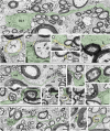
Oligodendrocytes respond to demyelination by contacting naked axons and engulfing them in their cytoplasm, resulting in restricted remyelination. In this montage, a range of the early and late interactions of mature oligodendrocytes with demyelinated axons is seen. Early contact with demyelinated axons is seen in AF and H, with fine processes (arrows) beginning to contact and surround axons (denoted by “a”). The adult oligodendrocyte in B is connected to two axons with mature myelin sheaths (*, g-ratios 0.75 and 0.67) and has a process connected to a demyelinated axon (a). Demyelinated axons can be seen indented within the oligodendrocyte cytoplasm (D) and within oligodendrocytes that have both mature and remyelinated sheaths (E). A mature oligodendrocyte myelinates both thin (*, g = 0.89) and thick (*, g = 0.55) sheaths (F). Remyelination is confirmed by the presence of short, thin internodes (G). [Scale bars: 5 µm (C, Inset), 10 µm (A, B, and DH), and 20 µm (C).]
Fig. 6.
Fig. 6.
EM confirms the oligodendrocyte/demyelinated axon interaction. (A and B) Two examples of oligodendrocytes connected to mature myelin sheaths that have initiated early ensheathment of demyelinated axons (denoted by “a”). (Scale bars, 2 µm.)
Fig. 7.
Fig. 7.
Cartoon illustrating the proposed response of mature oligodendrocytes to adjacent demyelinated internodes. There is partial loss of internodes of this oligodendrocytes territory and retraction of processes. This is quickly followed by process reextension and remyelination, resulting in thin, short internodes, while the axon maintains its original myelin sheaths.

Similar articles

Cited by

References

    1. Duncan ID, Brower A, Kondo Y, Curlee JF, Jr, Schultz RD. Extensive remyelination of the CNS leads to functional recovery. Proc Natl Acad Sci USA. 2009;106:6832–6836, and correction (2009) 106:12208. - PMC - PubMed
    1. Franklin RJM, Ffrench-Constant C. Regenerating CNS myelin—From mechanisms to experimental medicines. Nat Rev Neurosci. 2017;18:753–769. - PubMed
    1. Prineas JW, Connell F. Remyelination in multiple sclerosis. Ann Neurol. 1979;5:22–31. - PubMed
    1. Raine CS, Wu E. Multiple sclerosis: Remyelination in acute lesions. J Neuropathol Exp Neurol. 1993;52:199–204. - PubMed
    1. Lassmann H, Brück W, Lucchinetti C, Rodriguez M. Remyelination in multiple sclerosis. Mult Scler. 1997;3:133–136. - PubMed

Publication types

MeSH terms