. 2019 Feb 5;93(4):e01656-18.
doi: 10.1128/JVI.01656-18. Print 2019 Feb 15.

Closing and Opening Holes in the Glycan Shield of HIV-1 Envelope Glycoprotein SOSIP Trimers Can Redirect the Neutralizing Antibody Response to the Newly Unmasked Epitopes

Affiliations

Closing and Opening Holes in the Glycan Shield of HIV-1 Envelope Glycoprotein SOSIP Trimers Can Redirect the Neutralizing Antibody Response to the Newly Unmasked Epitopes

Rajesh P Ringe et al. J Virol. .

Abstract

In HIV-1 vaccine research, native-like, soluble envelope glycoprotein SOSIP trimers are widely used for immunizing animals. The epitopes of autologous neutralizing antibodies (NAbs) induced by the BG505 and B41 SOSIP trimers in rabbits and macaques have been mapped to a few holes in the glycan shields that cover most of the protein surfaces. For BG505 trimers, the dominant autologous NAb epitope in rabbits involves residues that line a cavity caused by the absence of a glycan at residue 241. Here, we blocked this epitope in BG505 SOSIPv4.1 trimer immunogens by knocking in an N-linked glycan at residue 241. We then opened holes elsewhere on the trimer by knocking out single N-linked glycans at residues 197, 234, 276, 332, and 355 and found that NAb responses induced by the 241-glycan-bearing BG505 trimers were frequently redirected to the newly opened sites. The strongest evidence for redirection of the NAb response to neoepitopes, through the opening and closing of glycan holes, was obtained from trimer immunogen groups with the highest occupancy of the N241 site. We also attempted to knock in the N289-glycan to block the sole autologous NAb epitope on the B41 SOSIP.v4.1 trimer. Although a retrospective analysis showed that the new N289-glycan site was substantially underoccupied, we found some evidence for redirection of the NAb response to a neoepitope when this site was knocked in and the N356-glycan site knocked out. In neither study, however, was redirection associated with increased neutralization of heterologous tier 2 viruses.IMPORTANCE Engineered SOSIP trimers mimic envelope-glycoprotein spikes, which stud the surface of HIV-1 particles and mediate viral entry into cells. When used for immunizing test animals, they elicit antibodies that neutralize resistant sequence-matched HIV-1 isolates. These neutralizing antibodies recognize epitopes in holes in the glycan shield that covers the trimer. Here, we added glycans to block the most immunogenic neutralization epitopes on BG505 and B41 SOSIP trimers. In addition, we removed selected other glycans to open new holes that might expose new immunogenic epitopes. We immunized rabbits with the various glycan-modified trimers and then dissected the specificities of the antibody responses. Thus, in principle, the antibody response might be diverted from one site to a more cross-reactive one, which would help in the induction of broadly neutralizing antibodies by HIV-1 vaccines based on envelope glycoproteins.

Keywords: Env trimer; HIV-1; SOSIP; antibodies; epitope; glycan hole; immunization; neutralization.


Figures

FIG 1
FIG 1
Neutralization of the parental BG505.T332N and immunogen-autologous viruses. In each panel, the ID50 values (y axis) for sera from individual rabbits are arranged by BG505 SOSIP.v.4.1 trimer immunogen group (see Results). The median value in each group is indicated by the horizontal bar. Only sera with ID50 values of >100 against the autologous or parental virus could be mapped precisely against mutant pseudoviruses (see subsequent figures). (A) Neutralization of the mutant pseudoviruses that are sequence matched (autologous) to the SOSIP trimer immunogens with respect to the glycan-KI and glycan-KO changes. The asterisk for group 4 signifies that the N241-KI + N197-KO double mutant virus was noninfectious; the data shown are for the N197-KO mutant. (B) Neutralization of the parental BG505.T332N pseudovirus.
FIG 2
FIG 2
Mutations affecting neutralization by group 1 sera (parental BG505 SOSIP.v4.1 trimer immunogen). The layout of this figure applies to each of Fig. 2 to 9 and 12 to 15, with the exceptions noted in those legends. (A) A surface-rendered model of the BG505 SOSIP.v4.1 trimer seen from the side, in two different orientations, with the apex up (top images) or viewed from above (lower image). In the subsequent figures, the orientation that most clearly displays the relevant glycan mutation is used. The peptidic surface is colored light blue on one of the protomers and gray on the other two (on some views not all three protomers are visible). The glycans are colored light green. The model is based on PDB entry 5V8M. (B) The data shown are the ID50 values against the parental BG505.T332N virus and the RID50 values (%) for neutralization of the indicated mutant pseudoviruses by the rabbit sera listed on the left. The RID50 value is equal to [(ID50 against mutant)/(ID50 against parental)] × 100%; hence, by definition it is 100% for the parental pseudovirus. The RID50 cells are color coded as outlined in the key above the panel. Intermediate and strong neutralization resistance is shown in yellow or red, respectively, while increased neutralization sensitivity is in blue. The Env mutations are indicated in the top row, and the complete sequence changes in the mutants are listed in Tables 1 and 2. RID50 values were only determined when the ID50 values against the parental or autologous pseudovirus were >100. KI, glycan knock in; KO, glycan knockout. RID50 values of >10 are rounded off to two significant digits and those of <10 to integers in Fig. 2 to 9 and Fig. 12 to 15.
FIG 3
FIG 3
Mutations affecting neutralization by group 2 sera (BG505 SOSIP.v4.1 + N241-KI trimer immunogen). The layout in panel A is analogous to that of Fig. 2A, except that residue S241 (left) and the glycan knocked in at residue 241 (right) are both colored yellow on the light blue protomer. The image on the left shows the hole that the new glycan blocks. The layout of panel B is analogous to that of Fig. 2B, where the color code is explained.
FIG 4
FIG 4
Mutations affecting neutralization by group 3 sera (BG505 SOSIP.v4.1 + N241-KI + N130-KI trimer immunogen). The color coding of the protomers and glycans of the trimers, seen from above, in panel A is the same as that in Fig. 2A, except that residue 130 (left) and the glycan knocked in at residue 130 (right) are both colored yellow. The image on the left shows the hole that the new glycan blocks. Note that the glycan knocked in at residue 241 is not visible, since the trimer is viewed from above. The layout of panel B is analogous to that of Fig. 2B, where the color code is explained.
FIG 5
FIG 5
Mutations affecting neutralization by group 4 sera (BG505 SOSIP.v4.1 + N241-KI + N197-KO trimer immunogen). The layout in panel A is analogous to that of Fig. 2A, except that the glycan knocked in at residue 241 is colored yellow on both images. The glycan normally present at residue 197 (left) and residue 197 itself (right) are both colored red to illustrate the hole created by the N197-KO mutation. The N355 glycan (light green) partly obscures the view of the N241 glycan, as in Fig. and . The layout of panel B is analogous to that of Fig. 2B, where the color code is explained. Note that the immunogen-matched N241-KI + N197-KO double mutant virus was noninfectious.
FIG 6
FIG 6
Mutations affecting neutralization by group 5 sera (BG505 SOSIP.v4.1 + N241-KI + N234-KO trimer immunogen). The layout in panel A is analogous to that of Fig. 5A. The glycan knocked in at residue 241 is colored yellow on both images, and the glycan knocked out at residue 234 (left) and residue 234 itself (right) are both colored red to illustrate the hole created by the N234-KO mutation. The layout of panel B is analogous to that of Fig. 2B, where the color code is explained.
FIG 7
FIG 7
Mutations affecting neutralization by group 6 sera (BG505 SOSIP.v4.1 + N241-KI + N276-KO trimer immunogen). The layout in panel A is analogous to that of Fig. 5A. To illustrate the hole created by the N276-KO mutation (i.e., by the T278A substitution), the glycan knocked in at residue 241 is colored yellow on both images and the glycan knocked out at residue 276 (left) and residue N276 itself (right) are both colored red. The layout of panel B is analogous to that of Fig. 2B, where the color code is explained. The four cells to the right for r2238-6 are open because the ID50 value against the parental virus was <100 and, hence, too low for precise mapping.
FIG 8
FIG 8
Mutations affecting neutralization by group 7 sera (BG505 SOSIP.v4.1 + N241-KI + N332-KO trimer immunogen). The layout in panel A is analogous to that of Fig. 5A. To illustrate the hole created by the N332-KO mutation, the glycan knocked in at residue 241 is colored yellow on both images and the glycan knocked-out at residue 332 (left) and residue 332 itself (right image) are both colored red. The layout of panel B is comparable to that of Fig. 2B, where the color code is explained. Four cells are open for r2243-7 because the ID50 value against the parental virus was <100 and, hence, was too low for precise mapping.
FIG 9
FIG 9
Mutations affecting neutralization by group 8 sera (BG505 SOSIP.v4.1 + N241-KI + N355-KO trimer immunogen). The layout in panel A is analogous to that of Fig. 5A. To illustrate the hole created by the N355-KO mutation, the glycan knocked in at residue 241 is colored yellow on both images and the glycan knocked out at residue 355 (left image) and residue 355 itself (right image) are both colored red. The N355 glycan partly covers the N241 glycan in this view. The layout of panel B is analogous to that of Fig. 2B, where the color code is explained.
FIG 10
FIG 10
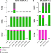
Proteomics-based quantification of glycan occupancy at glycan-KI sites on SOSIP trimers. (A) The three diagrams show the proportions (y axes) of the N130, N241, or N332 sites occupied by oligomannose glycans (green) or complex-type glycans (pink) or not occupied (gray) for the BG505 SOSIP.v4.1 trimer immunogens listed on the category axis. (B) As for panel A, except the data are for the N289, N332, or N356 sites on the B41 SOSIP.v4.1 trimer immunogens. For both panels, the error bars represent SEM for multiple peptides per site. N/A, not applicable (i.e., the site was naturally absent or had been knocked out).
FIG 11
FIG 11
Neutralization of the parental B41.R315Q and immunogen-autologous viruses. In each panel, the ID50 values (y axis) for sera from individual rabbits are arranged by B41 SOSIP.v4.1 trimer immunogen group (see Results). The median value in each group is indicated by the horizontal bar. Only sera with ID50 values of >100 against the autologous or parental virus could be mapped with any precision against mutant pseudoviruses (see subsequent figures). (A) Neutralization of the mutant pseudoviruses that are sequence matched (autologous) to the SOSIP trimer immunogens with respect to the glycan-KI and glycan-KO changes. (B) Neutralization of the parental B41.R315Q pseudovirus.
FIG 12
FIG 12
Mutations affecting neutralization by group 9 sera (parental B41 SOSIP.v4.1 trimer immunogen). (A) Surface-rendered model of the B41 SOSIP.v4.1 trimer seen from the side in two different orientations, with the apex up (top) or (lower) viewed from above. The peptidic surface is colored light blue on one of the protomers and gray on the other two (on some views not all the protomers are visible). The glycans are colored light green on all three. The three-dimensional model of the B41 SOSIP.v4.1 trimer was based on an unpublished crystal structure of B41 SOSIP.664 at 3.5 Å. The layout of panel B is analogous to that of Fig. 2B, where the color code is explained, except that the parental virus is B41.R315Q.
FIG 13
FIG 13
Mutations affecting neutralization by group 10 sera (B41 SOSIP.v4.1 + N289-KI trimer immunogen). The layout in panel A is analogous to that of Fig. 2A, except that residue N289 (left) and the glycan knocked in at residue 289 (right) are both colored yellow on the light blue protomer. The image on the left shows the hole that the new glycan blocks. The E290 and Q344 residues partly cover N289. The layout of panel B is analogous to that of Fig. 2B, where the color code is explained.
FIG 14
FIG 14
Mutations affecting neutralization by group 11 sera (B41 SOSIP.v4.1 + N289-KI + N332-KO trimer immunogen). The layout in panel A is analogous to that of Fig. 5A. To illustrate the hole created by the N332-KO mutation, the glycan knocked in at residue 289 is colored yellow on both images and the glycan knocked out at residue 332 (left image) and residue 332 itself (right image) are both colored red. The layout of panel B is analogous to that of Fig. 2B, where the color code is explained.
FIG 15
FIG 15
Mutations affecting neutralization by group 12 sera (B41 SOSIP.v4.1 + N289-KI + N356-KO trimer immunogen). The layout in panel A is analogous to that of Fig. 5A. To illustrate the hole created by the N356-KO mutation, the glycan knocked in at residue 289 is colored yellow in both images and the glycan knocked out at residue 356 is colored red in both images. The N356 glycan appears small because only its GlcNac component is shown. The layout of panel B is analogous to that of Fig. 2B, where the color code is explained (45).
FIG A1
FIG A1
Comparison of neutralization of autologous and parental pseudoviruses. In each of the groups with nonparental BG505 SOSIP.v4.1 immunogens (groups 2 to 8), the ID50 values for neutralization of autologous and parental pseudoviruses are compared. Above each group designation (group number and mutations in the immunogen), a short line represents individual rabbits and connects the ID50 value against the autologous (or near-autologous) virus at its left end with the corresponding value against the parental virus at its right end. In group 2 the lines for two negative sera are superimposed (ID50 of ≤20). Except in groups 2 and 3, i.e., those groups without glycan-KO mutations in the immunogen, many of the lines have downward slopes, indicating redirection to epitopes unmasked by the mutations. This tendency was significant in group 5.
FIG A3
FIG A3
General evidence for redirection to glycan-KO neoepitopes. The ID50 values against the immunogen-matching glycan-KO mutant pseudoviruses (blue symbols, left column; n = 45) were compared with those against non-immunogen-matching glycan-KO mutants (red symbols, right column; n = 315). The matching serum-virus combinations had significantly higher ID50 values than the nonmatching ones (P < 0.0001).
FIG A2
FIG A2
Neutralization of tier 1A viruses by rabbit sera. The ID50 values against the tier 1A pseudoviruses MN.3 (left) and MW965.26 (right) are depicted above the respective group designations (group number and mutations in the immunogen) for the BG505 SOSIP.v4.1 immunization groups 1 to 8 (A) and the B41 SOSIP.v4.1.R315Q immunization groups 9 to 12 (B).
FIG A4
FIG A4
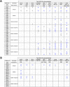
Heterologous neutralization of tier 2 viruses by rabbit sera. The sera from BG505 groups 1 to 8 (A) and B41 groups 9 to 12 (B) were tested against a standard global panel of 9 tier 2 viruses (61). The titers (ID50 values) were determined in the Tzm-bl assay. All background-corrected ID50 values of >40 (see Materials and Methods) are marked in blue and boldface, values in the range of 21 to 40 are in black, and values of ≤20, regarded as negative, are represented by a dash. The data for the BG505 study were determined at DUMC. For the B41 study, the sera were first tested at DUMC and then at the AMC. Sera from the three rabbits with the highest responses (2257-9, 2267-11, and 2268-11) were also retested at DUMC. The concordance between the two centers in positive and negative results was >90%. Thus, values in panel B are means from 2 to 3 independent assays. (For intra-assay replicates and controls, see Materials and Methods). The absence (−) or presence (+) of glycan sites that correspond to some sites that are mutated in the BG505 and B41 SOSIP.v4.1 trimer immunogen groups is indicated under the virus designations. The BG505 SOSIP.v4.1 trimer immunogens for groups 2 to 8 all contained the N241-KI mutation, and their similarity with 8 of the 9 viruses (all except 25710) at that site is not listed; likewise, all viruses have N197, which was knocked out only in the immunogen of group 4; the similarity at residue 197 with all the others also is not listed. The glycans in the region of residues 355 to 360 are shifted slightly in relation to the HXB2 sequence, complicating comparisons at that site.
FIG A5
FIG A5
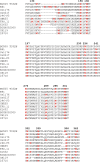
Alignment of sequences around mutated PNGS. The amino acid sequences of Env parental viruses BG505.T332N and B41.R315Q and nine tier 2 viruses in the global panel (Fig. A4) were aligned. Regions comprising sequons for N-linked glycosylation that were mutated in the immunogen constructs are shown. Asn residues in PNGSs are marked in red; HXB2-derived numbering of the Asn residues is given in boldface at the top.

References

    1. Escolano A, Dosenovic P, Nussenzweig MC. 2017. Progress toward active or passive HIV-1 vaccination. J Exp Med 214:3–16. doi:10.1084/jem.20161765. - DOI - PMC - PubMed
    1. Sanders RW, Moore JP. 2017. Native-like Env trimers as a platform for HIV-1 vaccine design. Immunol Rev 275:161–182. doi:10.1111/imr.12481. - DOI - PMC - PubMed
    1. Ward AB, Wilson IA. 2017. The HIV-1 envelope glycoprotein structure: nailing down a moving target. Immunol Rev 275:21–32. doi:10.1111/imr.12507. - DOI - PMC - PubMed
    1. Bonsignori M, Liao HX, Gao F, Williams WB, Alam SM, Montefiori DC, Haynes BF. 2017. Antibody-virus co-evolution in HIV infection: paths for HIV vaccine development. Immunol Rev 275:145–160. doi:10.1111/imr.12509. - DOI - PMC - PubMed
    1. Klein F, Mouquet H, Dosenovic P, Scheid JF, Scharf L, Nussenzweig MC. 2013. Antibodies in HIV-1 vaccine development and therapy. Science 341:1199–1204. doi:10.1126/science.1241144. - DOI - PMC - PubMed

Publication types

LinkOut - more resources