Review
. 2018 Nov 28;10(12):471.
doi: 10.3390/cancers10120471.

Chemoresistance and the Self-Maintaining Tumor Microenvironment

Affiliations
Review

Chemoresistance and the Self-Maintaining Tumor Microenvironment

Gulcen Yeldag et al. Cancers (Basel). .

Abstract

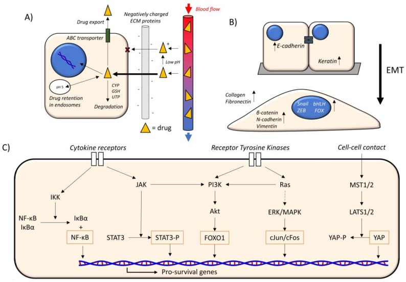
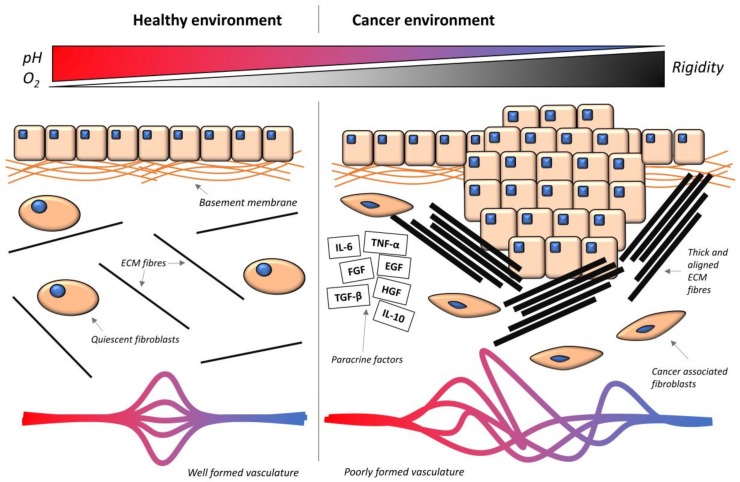
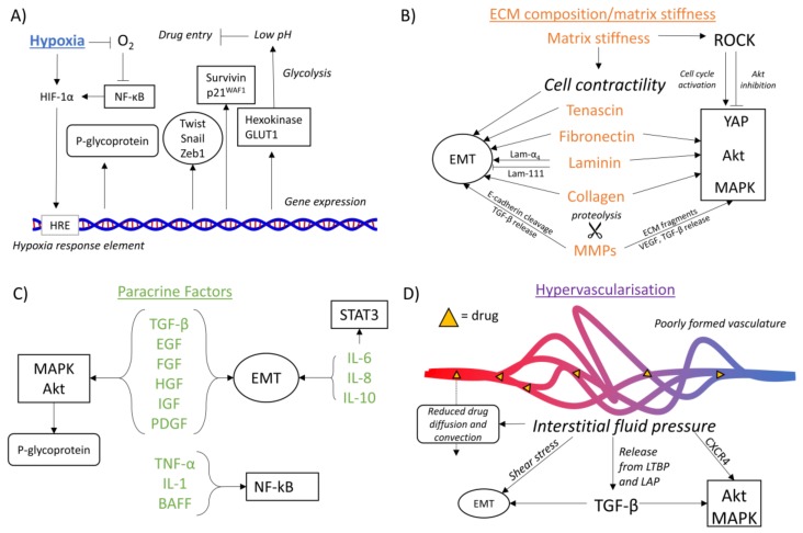
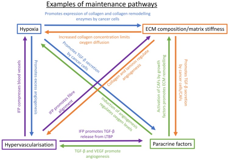
The progression of cancer is associated with alterations in the tumor microenvironment, including changes in extracellular matrix (ECM) composition, matrix rigidity, hypervascularization, hypoxia, and paracrine factors. One key malignant phenotype of cancer cells is their ability to resist chemotherapeutics, and elements of the ECM can promote chemoresistance in cancer cells through a variety of signaling pathways, inducing changes in gene expression and protein activity that allow resistance. Furthermore, the ECM is maintained as an environment that facilitates chemoresistance, since its constitution modulates the phenotype of cancer-associated cells, which themselves affect the microenvironment. In this review, we discuss how the properties of the tumor microenvironment promote chemoresistance in cancer cells, and the interplay between these external stimuli. We focus on both the response of cancer cells to the external environment, as well as the maintenance of the external environment, and how a chemoresistant phenotype emerges from the complex signaling network present.

Keywords: ECM; cancer stem cells; chemoresistance; fibrosis; hypervascularization; hypoxia; mechanosignaling; paracrine.


Conflict of interest statement

The authors declare no conflict of interest.

Figures

Figure 1
Figure 1
Mechanisms of chemoresistance. (A) Drug availability. Drugs (yellow triangles) move from blood vessels (right) to cells. A low pH can promote charged drugs that interact with negatively-charged extracellular matrix (ECM) components (grey), slowing their transit. Charged drugs are also hindered in their ability to cross the hydrophobic plasma membrane. Inside the cell, drugs can move to the nucleus to achieve their cytotoxic effects. Alternatively, drugs can be transported out of the cell by ABC transporters, and degraded by the cytochrome P450 system (CYP), glutathione-S-transferase (GSH) superfamily or uridine diphospho-glucuronosyltransferase (UGT) superfamily. Drugs also enter endosomes, and charged drugs are retained more within these acidic vesicles. (B) EMT. The epithelial–mesenchymal transition (EMT) involves the loss of cell–cell adhesion from the epithelial phenotype (above) to the mesenchymal phenotype (below). E-cadherin and keratin are more abundant in the epithelial phenotype. β-catenin, N-cadherin, and vimentin, and the transcription factor families Snail, ZEB, bHLH, and FOX, are more abundant in the mesenchymal phenotype. Collagen and fibronectin secretion is also increased in the mesenchymal phenotype. (C) Oncogenic signaling pathways. The main oncogenic signaling pathways that lead to the expression of pro-survival genes. Cytokine receptors promote NF-κB and STAT signaling, receptor tyrosine kinases promote PI3K and MAPK signaling, and cell–cell contact inhibits YAP signaling (Hippo pathway). The orange outline indicates a component that is involved directly in promoting transcription. IKK = IκB kinase, NF-κB = nuclear factor kappa-light-chain-enhancer of activated B cells, IκBα = inhibitor of kappa B, JAK = Janus kinase, STAT3 = Signal transducer and activator of transcription 3, STAT3-P = phosphorylated STAT3, PI3K = Phosphatidylinositol-4,5-bisphosphate 3-kinase, Akt = protein kinase B, FOXO1 = forkhead box protein O1, ERK/MAPK = mitogen-activated protein kinase, MST1/2 = mammalian ste20 homolog 1/2, LATS1/2 = large tumor suppressor kinase 1/2, YAP = Yes-associated protein, YAP-P = phosphorylated YAP. Dotted arrow = multiple reactions.
Figure 2
Figure 2
Comparison between the healthy and the cancer microenvironment. In the healthy environment, epithelial cells are separated from the stroma below by the basement membrane. In the stroma, fibroblasts show their quiescent phenotype, and there is a low abundance of ECM fibers (black), and therefore reduced rigidity. The vasculature is highly organized, and there is a high level of oxygen and a neutral pH. In the cancer environment, excess cell growth leads to breaching of the basement membrane, and cells are in contact with the stroma. Cancer-associated fibroblasts (CAFs) become activated, and there is a high abundance of ECM fibers and growth factors (black outlined rectangles). TNF-α = tumor necrosis factor alpha, IL-6 = interleukin 6, IL-10 = interleukin 10, EGF = epidermal growth factor, FGF = fibroblast growth factor, HGF = hepatic growth factor, TGF-β = transforming growth factor beta. The vasculature is excessive but poorly organized, and oxygen and pH are reduced. Stromal rigidity is increased.
Figure 3
Figure 3
Effects of the microenvironment on mechanisms of chemoresistance. (A) Hypoxia. A lack of oxygen leads to the stabilization of HIF-1α (hypoxia inducible factor 1 alpha), either directly or through NF-κB-mediated (nuclear factor kappa-light-chain-enhancer of activated B cells) gene expression. HIF-1α binds to the hypoxia response element (HRE), upregulating P-glycoprotein (drug availability), the transcription factors Twist, Snail, and Zeb1 (EMT) and survivin and p21WAF1 (cyclin-dependent kinase inhibitor 1) (oncogenic signaling). Also upregulated are hexokinase and GLUT1 (glucose transporter 1), which promote glycolysis, leading to a reduced external pH and inhibition of drug entry. (B) ECM composition and matrix stiffness. The ECM proteins tenascin, fibronectin, laminin, and collagen all affect the EMT (epithelial–mesenchymal transition), although different laminin α chains have different effects. Fibronectin, laminin, and collagen also modulate the oncogenic signaling pathways of YAP, Akt, and MAPK. MMPs (matrix metalloproteinases) act to release paracrine factors such as TGF-β, which regulates both EMT and oncogenic pathways. In addition, MMPs can cleave E-cadherin to promote EMT and ECM fragments to promote oncogenic pathways. Matrix stiffness promotes cell contractility, which leads to EMT, as well as promoting Rho-associated coiled coil-containing protein kinase (ROCK) activation, which has differing effects on oncogenic pathways. (C) Paracrine factors. The factors TGF-β, EGF, FGF, HGF, insulin-like growth factor (IGF), and platelet derived growth factor (PDGF) both promote EMT, and the MAPK and Akt pathways. MAPK and Akt also activate P-glycoprotein. Interleukins 6 and 8 promote STAT3 signaling and EMT. TNF-α, IL-1 and B cell-activating factor (BAFF) promoter NF-κB signaling. (D) Hypervascularization. The interstitial fluid pressure generated by hypervascularization can promote EMT through shear stress, oncogenic signaling pathways through C-X-C chemokine receptor type 4 (CXCR4) activation, and TGF-β release, which promotes EMT and oncogenic pathways. Interstitial fluid pressure reduces drug diffusion and convection. In all cases, rounded rectangle = drug availability, circle = EMT, rectangle = oncogenic signaling pathways.
Figure 4
Figure 4
Examples of environment maintenance pathways. Blue = hypoxia-induced. Orange = ECM composition/matrix stiffness induced. Green = Paracrine factor induced. Purple = hypervascularization induced. TGF-β = transforming growth factor beta, IFP = interstitial fluid pressure, CAFs = cancer-associated fibroblasts, LTBP = latent TGF-β-binding protein, VEGF = vascular endothelial growth factor, ECM = extracellular matrix.

Similar articles

Cited by

References

    1. Zheng H. The molecular mechanisms of chemoresistance in cancers. Oncotarget. 2017;8:59950–59964. doi: 10.18632/oncotarget.19048. - DOI - PMC - PubMed
    1. Senthebane D.A., Rowe A., Thomford N.E., Shipanga H., Munro D., Mazeedi M., Almazyadi H.A.M., Kallmeyer K., Dandara C., Pepper M.S., et al. The Role of Tumor Microenvironment in Chemoresistance: To Survive, Keep Your Enemies Closer. Int. J. Mol. Sci. 2017;18:1586. doi: 10.3390/ijms18071586. - DOI - PMC - PubMed
    1. Cox T.R., Erler J.T. Remodeling and homeostasis of the extracellular matrix: Implications for fibrotic diseases and cancer. Dis. Model. Mech. 2011;4:165–178. doi: 10.1242/dmm.004077. - DOI - PMC - PubMed
    1. Zhang X., Nie D., Chakrabarty S. Growth factors in tumor microenvironment. Front. Biosci. 2012;15:151–165. doi: 10.2741/3612. - DOI - PMC - PubMed
    1. Petrova V., Annicchiarico-Petruzzelli M., Melino G., Amelio I. The hypoxic tumour microenvironment. Oncogenesis. 2018;7:10. doi: 10.1038/s41389-017-0011-9. - DOI - PMC - PubMed