Review
. 2019 Apr;16(4):256-268.
doi: 10.1038/s41571-018-0135-7.

Clinical potential of mass spectrometry-based proteogenomics

Affiliations
Review

Clinical potential of mass spectrometry-based proteogenomics

Bing Zhang et al. Nat Rev Clin Oncol. 2019 Apr.

Abstract

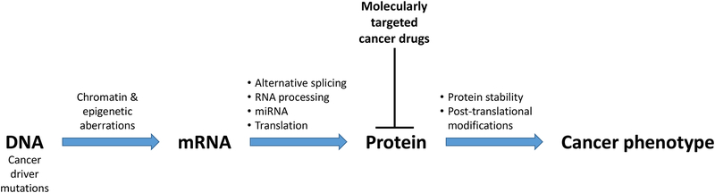
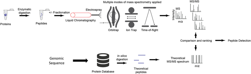
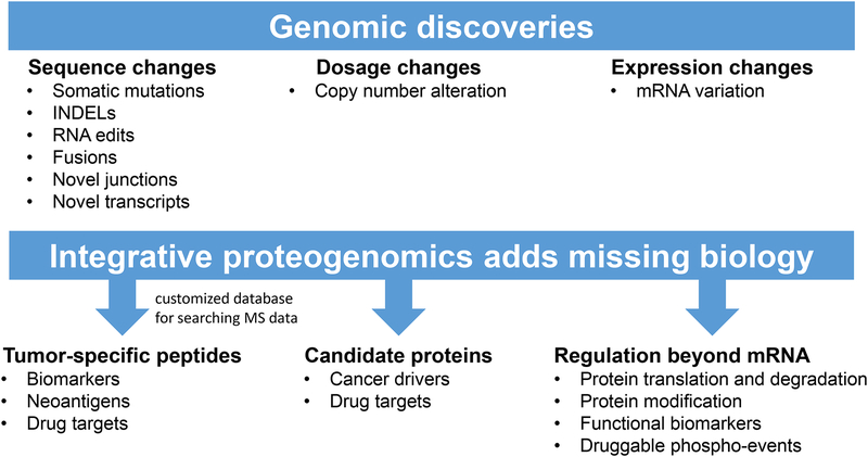
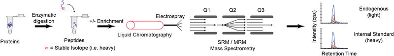
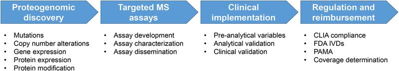
Cancer genomics research aims to advance personalized oncology by finding and targeting specific genetic alterations associated with cancers. In genome-driven oncology, treatments are selected for individual patients on the basis of the findings of tumour genome sequencing. This personalized approach has prolonged the survival of subsets of patients with cancer. However, many patients do not respond to the predicted therapies based on the genomic profiles of their tumours. Furthermore, studies pairing genomic and proteomic analyses of samples from the same tumours have shown that the proteome contains novel information that cannot be discerned through genomic analysis alone. This observation has led to the concept of proteogenomics, in which both types of data are leveraged for a more complete view of tumour biology that might enable patients to be more successfully matched to effective treatments than they would using genomics alone. In this Perspective, we discuss the added value of proteogenomics over the current genome-driven approach to the clinical characterization of cancers and summarize current efforts to incorporate targeted proteomic measurements based on selected/multiple reaction monitoring (SRM/MRM) mass spectrometry into the clinical laboratory to facilitate clinical proteogenomics.


Figures

Figure 1.
Figure 1.. Many processes downstream of the genome affect the cancer phenotype.
Proteins execute the genome to control tumor phenotype, and proteins are most frequently targeted in precision oncology.
Figure 2.
Figure 2.. Untargeted (i.e. “shotgun”) discovery proteomics.
In untargeted (i.e. ‘shotgun’) proteomics, proteins are converted to peptides through proteolytic digestion (typically using trypsin). The peptides are fractionated by liquid chromatography and introduced to a mass spectrometer through electrospray ionization. For detection of low abundance proteins, an additional fractionation or enrichment step may be performed. Multiple modes of mass spectrometry are used, depending on available instrumentation and the design of the experiment. For example, an Orbitrap instrument uses image current detection to measure ions oscillating around the central electrode. Fourier transformation converts the signal from the time domain to the frequency domain, producing the mass spectrum. The Orbitrap is capable of high resolution and mass accuracy with a trade-off of relatively longer acquisition times. A linear ion trap uses static and RF (radiofrequency) fields to confine ions within the trap. The RF voltage is adjusted to confine/eliminate desired ions for detection. The ion trap is fast, but the spectra have relatively lower resolution and mass accuracy. Finally, a time-of-flight instrument measures the drift time of ions as they pass through a field-free region. The drift time is proportional to the mass-to-charge ratio. Time-of-flight acquisition is relatively fast, and resolution varies depending on instrument model. Regardless of instrument type, the most common structure of an untargeted proteomics dataset is data-dependent, i.e. it consists of a survey MS scan followed by MS/MS spectra of individual ions (peptides). The MS/MS spectra are searched against a database of known sequences to detect the peptide sequence.
Figure 3.
Figure 3.. Integrative proteogenomic data analysis adds missing biology.
Genomic data identify somatic changes in tumors, and enable the generation of custom databases for searching MS data. Proteomic data confirm the translation of genomic results and also uncover additional biology (e.g. post-translational modifications).
Figure 4.
Figure 4.. Targeted MS-based assays.
In targeted proteomics, proteins are converted to peptides through enzymatic (typically trypsin) digestion. The peptides are separated by liquid chromatography and introduced to a mass spectrometer through electrospray ionization. For low abundance proteins, a fractionation or enrichment step may be performed, such as immunoaffinity enrichment (e.g. immuno-MRM assay). Stable isotope-labeled peptides are added as internal standards and SRM/MRM analysis uses a triple quadrupole mass spectrometer (QQQ). The first and third quadrupoles (Q1, Q3) are used as mass filters separated by a collision cell for fragmentation (Q2). Q1 is tuned to pass the m/z of the target ion (i.e. the precursor ion), while filtering all other ions. The targeted precursor ion is fragmented by collision-induced dissociation in Q2, producing fragment ions (i.e. product ions). Q3 is used as a mass filter to pass a single analyte-specific product ion for detection. The combination of a specific precursor / product ion pair is termed a “transition.” The data output is a peak area ratio of the endogenous analyte peptide relative to the internal standard. By monitoring transitions, sensitivity is improved over untargeted methods by removing chemical noise and increasing the signal-to-noise ratio for the analyte. Specificity is also high by incorporating multiple levels of isolation and choosing highly specific transitions. Multiplexing is achieved by sequentially stepping through a list of transitions. Acquisition of multiple transitions is typically performed on a timescale that is tens to hundreds of times faster than the chromatographic peak widths of eluting peptides, enabling multiple points to be measured across a peak. The measurement of up to several hundred peptides are readily multiplexed into a single analytical run.
Figure 5.
Figure 5.. Workflow for clinical implementation of proteogenomics.

Similar articles

Cited by

References

    1. Vogelstein B et al. Cancer genome landscapes. Science 339, 1546–1558, doi:10.1126/science.1235122 (2013). - DOI - PMC - PubMed
    1. Simon R Critical Review of Umbrella, Basket, and Platform Designs for Oncology Clinical Trials. Clin Pharmacol Ther 102, 934–941, doi:10.1002/cpt.814 (2017). - DOI - PubMed
    1. Maitland ML & Schilsky RL Clinical trials in the era of personalized oncology. CA Cancer J Clin 61, 365–381, doi:10.3322/caac.20135 (2011). - DOI - PMC - PubMed
    1. Subbiah V & Kurzrock R Debunking the Delusion That Precision Oncology Is an Illusion. Oncologist 22, 881–882, doi:10.1634/theoncologist.2017-0040 (2017). - DOI - PMC - PubMed
    1. Hyman DM, Taylor BS & Baselga J Implementing Genome-Driven Oncology. Cell 168, 584–599, doi:10.1016/j.cell.2016.12.015 (2017). - DOI - PMC - PubMed

Publication types

Substances