. 2018 Nov;63(3):169-174.
doi: 10.3164/jcbn.17-133. Epub 2018 Jul 12.

Heme oxygenase-1 prevents murine intestinal inflammation

Affiliations

Heme oxygenase-1 prevents murine intestinal inflammation

Tomohisa Takagi et al. J Clin Biochem Nutr. 2018 Nov.

Abstract

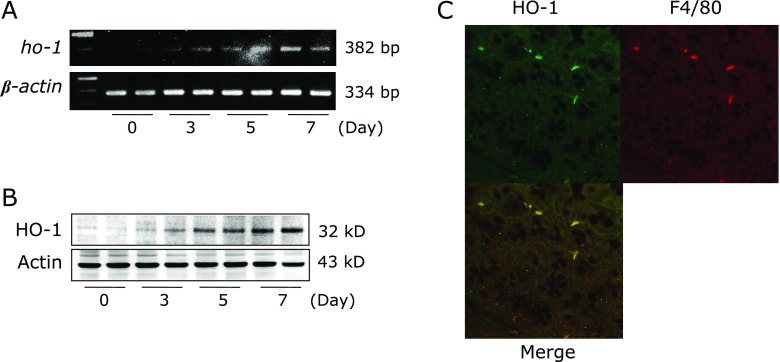
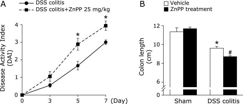
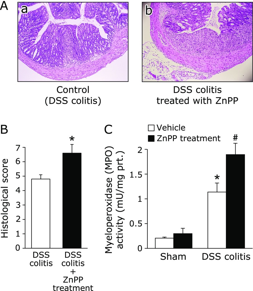
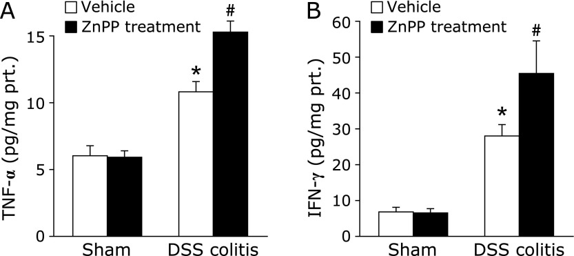
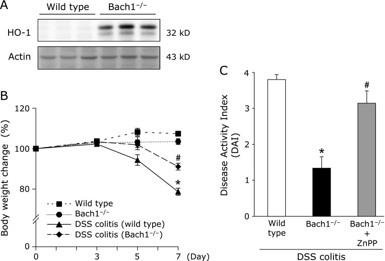
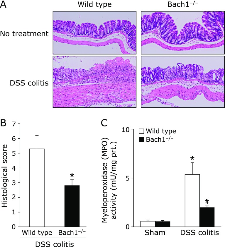
Heme oxygenases (HOs) are rate-limiting enzymes catabolizing heme to biliverdin, ferrous iron, and carbon monoxide, and of the three HO isoforms identified, HO-1 plays a protective role against inflammatory processes. In this study, we investigated the possible role of HO-1 in intestinal inflammation. Acute colitis was induced in male C57BL/6 (wild-type) and homozygous BTB and CNC homolog 1 (Bach1)-deficient mice, which show high HO-1 expression in the colonic mucosa, using dextran sodium sulfate. The disease activity index, myeloperoxidase activity, and inflammatory cytokines in the colonic mucosa were evaluated 7 days after dextran sodium sulfate-dependent colitis induction. We also evaluated the impact of HO-1 inhibition using zinc protoporphyrin IX (25 mg/kg i.p., daily). After dextran sodium sulfate administration, HO-1 mRNA and protein expression increased in a time-dependent manner. Disease activity index score, myeloperoxidase activity, and colonic production of TNF-α and IFN-γ were increased after dextran sodium sulfate administration, and co-administration of zinc protoporphyrin IX enhanced their increase. In addition, disease activity index in Bach1-deficient was significantly lower after dextran sodium sulfate administration than that in wild type mice. These results indicate that HO-1 plays a protective role against dextran sodium sulfate-induced intestinal inflammation, possibly by regulating pro-inflammatory cytokines in intestinal tissues.

Keywords: BTB and CNC homolog 1 (Bach1); dextran sodium sulfate (DSS)-induced colitis; heme oxygenase-1 (HO-1).


Conflict of interest statement

No potential conflicts of interest were disclosed.

Figures

Fig. 1
Fig. 1
Effect of zinc protoporphyrin (ZnPP) on DSS-induced colitis. To analyze HO-1 expression in colonic mucosa, time-dependent expression of ho-1 mRNA in colonic mucosal tissue after DSS administration was determined using RT-PCR (A). Time-dependent expression of HO-1 protein in colonic mucosa after DSS administration was determined by western blotting (B). The results representative of 3 separate experiments. Each treatment condition after DSS treatment is represented by 2 lanes. (C) Colonic Localization of HO-1 expression was analyzed by immunofluorescence staining and visualized under a laser scanning confocal microscope. Upper left: HO-1 stained with anti-rabbit Alexa 594 secondary antibody; Upper right: F4/80 stained with anti-rat Alexa 488 secondary antibody; Below: merged image of upper images. A representative image from three separate experiments is shown.
Fig. 2
Fig. 2
Effects of HO-1 inhibition on dextran sulfate sodium (DSS)-induced colitis. (A) The effect of ZnPP on the disease activity index during the development of dextran sulfate sodium (DSS)-induced colitis in mice. Each value indicates the mean ± SE for 8 mice. *p<0.05 compared with control mice receiving 2% DSS solution alone. (B) The effect of ZnPP on the total length of the colon after DSS administration. Each value indicates the mean ± SE for 8 mice. *p<0.05 compared with sham mice receiving vehicle, #p<0.05 compared with control mice receiving 2% DSS solution alone.
Fig. 3
Fig. 3
Deterioration of colitis by HO-1 inhibition. (A) The histological appearance of colonic tissue in mice with DSS induced-colitis receiving vehicle (a) and those treated with ZnPP (b). Loss and shortening of crypts, mucosal erosions, inflammatory cell infiltration, and goblet cell depletion are seen in (a). In (b), larger erosions are associated with much inflammatory cell infiltration. Hematoxylin and eosin staining. Magnification, ×40. (B) Histological score was evaluated as described in Materials and Methods. The data represent the mean ± SE of 5 mice. *p<0.05 compared with mice with DSS induced-colitis receiving vehicle. (C) Effect of ZnPP on neutrophil accumulation expressed as myeloperoxidase (MPO) activity. Each value indicates the mean ± SE for 7 mice. *p<0.05 compared with mice receiving vehicle, #p<0.05 compared with control mice receiving 2% DSS solution alone.
Fig. 4
Fig. 4
Effect of ZnPP on tumor necrosis factor (TNF)-α (A) and interferon (IFN)-γ (B) in the colonic mucosa of mice. Each value indicates the mean ± SE for 7 mice. *p<0.05 compared with mice receiving the vehicle, #p<0.05 compared with control mice receiving 2% DSS solution alone.
Fig. 5
Fig. 5
Bach1 deficiency ameliorated DSS-induced colitis. (A) HO-1 protein expression in the colonic mucosa of Bach1-deficient mice was determined by western blotting. Results are representative of 3 separate experiments. (B) The degree of body weight loss during DSS treatment. Each value indicates the mean ± SE for 6 mice. *p<0.05 compared with wild type mice receiving vehicle, #p<0.05 compared with wild type mice receiving 2% DSS solution. (C) Disease activity index (DAI) on day 7 of DSS treatment. The effect of ZnPP on DAI during the development of DSS-induced colitis in Bach1-deficient mice was also evaluated. Each value indicates the mean ± SE for 5 mice. *p<0.05 compared with wild type mice receiving 2% DSS solution, #p<0.05 compared with Bach1-deficient mice receiving 2% DSS solution.
Fig. 6
Fig. 6
Bach1 deficiency ameliorated DSS-induced colitis. (A) The histological appearance of colonic tissue in wild type mice and Bach1-deficient mice without or with DSS induced-colitis. Hematoxylin and eosin staining. Magnification, ×40. (B) Histological score was evaluated as described in Materials and Methods. Data represent the mean ± SE of 5 mice. *p<0.05 compared to wild type mice with DSS induced-colitis. (C) Tissue-associated neutrophil accumulation in the colonic mucosa expressed as myeloperoxidase (MPO) activity. Each value indicates the mean ± SE for 7 mice. *p<0.05 compared with wild type mice without 2% DSS solution, #p<0.05 compared with wild type mice receiving 2% DSS solution.

References

    1. Kaplan GG, Ng SC. Understanding and preventing the global increase of inflammatory bowel disease. Gastroenterology. 2017;152:313–321. - PubMed
    1. Xavier RJ, Podolsky DK. Unravelling the pathogenesis of inflammatory bowel disease. Nature. 2007;448:427–434. - PubMed
    1. Alzoghaibi MA. Concepts of oxidative stress and antioxidant defense in Crohn’s disease. World J Gastroenterol. 2013;19:6540–6547. - PMC - PubMed
    1. Naito Y, Takagi T, Yoshikawa T. Neutrophil-dependent oxidative stress in ulcerative colitis. J Clin Biochem Nutr. 2007;41:18–26. - PMC - PubMed
    1. Takagi T, Naito Y, Yoshikawa T. Free radicals in inflammatory bowel disease. In: Naito Y, Suematsu M, Yoshikawa T, editors. Frontiers of Gastrointestinal Research; Free Radical Biology in Digestive Disease. Basel (Switzerland): Karger Publishers; 2011. pp. 128–136.