

Decreased Functional Connectivity in Insular Subregions in Depressive Episodes of Bipolar Disorder and Major Depressive Disorder
- PMID: 30487732
- PMCID: PMC6246657
- DOI: 10.3389/fnins.2018.00842
Decreased Functional Connectivity in Insular Subregions in Depressive Episodes of Bipolar Disorder and Major Depressive Disorder
Abstract
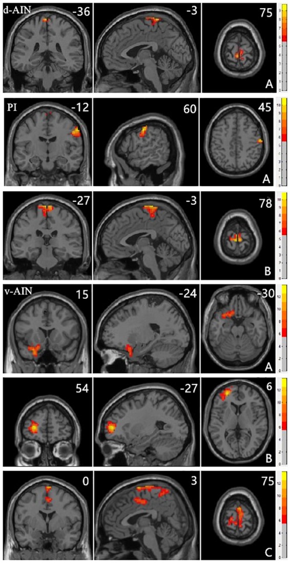
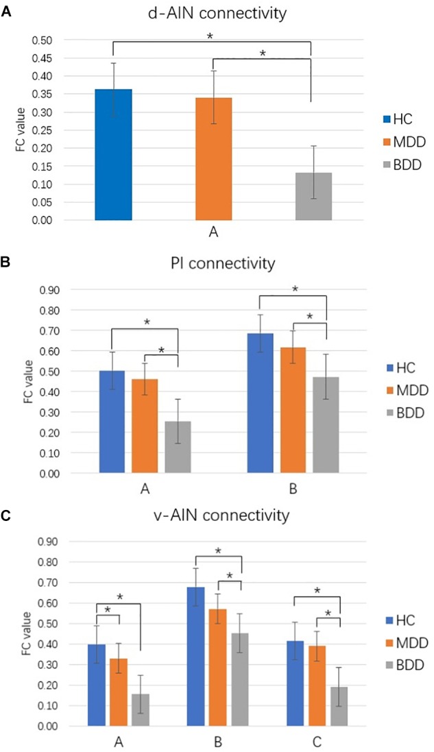
Objective: Clinically, it is very difficult to distinguish between major depressive disorder (MDD) and bipolar disorder (BD) in the period of depression. Increasing evidence shows that the insula plays an important role in depression. We aimed to compare the resting-state functional connectivity (rsFC) of insular subregions in patients with MDD and BD in depressive episodes (BDD), who had never experienced manic or hypomanic episodes when they were scanned to identify biomarkers for the identification of two diseases. Methods: We recruited 21 BDD patients, 40 MDD patients and 70 healthy controls (HC). Resting-state functional magnetic resonance imaging (rs-fMRI) was performed. BDD patients had never had manic or hypomanic episodes when they were scanned, and the diagnoses were determined by follow-up. We divided the insula into three parts including the ventral anterior insular cortex (v-AIN), dorsal anterior insular cortex (d-AIN), and posterior insula (PI). The insular-based rsFC was compared among the three groups, and an analysis of the correlation between the rsFC value and Hamilton depression and anxiety scales was carried out. Results: BDD and MDD patients demonstrated decreased rsFC from the v-AIN to the left superior/middle frontal gyrus compared with the HC group. Versus MDD and HC groups, BDD patients exhibited decreased rsFC from the v-AIN to the area in the left orbital frontal gyrus and left superior temporal gyrus (included temporal pole), from the PI to the right lateral postcentral gyrus and from all three insular subregions to the somatosensory and motor cortex. Meanwhile, a correlation between the rsFC value of the PI-right lateral postcentral gyrus and anxiety score was observed in patients. Conclusion: Our findings show BDD and MDD patients have similar decreases in insular connectivity in the dorsal lateral frontal regions, and BDD patients have specific decreased insular connectivity, especially in the somatosensory and motor cortex, which may be used as imaging evidence for clinical identification.
Keywords: bipolar; depressive; fMRI; functional connectivity; insula.
Figures
Similar articles
-
The Insular Subregions Topological Characteristics of Patients With Bipolar Depressive Disorder.Front Psychiatry. 2020 Apr 15;11:253. doi: 10.3389/fpsyt.2020.00253. eCollection 2020. Front Psychiatry. 2020. PMID: 32351411 Free PMC article.
-
Insula and amygdala resting-state functional connectivity differentiate bipolar from unipolar depression.Acta Psychiatr Scand. 2017 Jul;136(1):129-139. doi: 10.1111/acps.12724. Epub 2017 Mar 28. Acta Psychiatr Scand. 2017. PMID: 28369737 Free PMC article.
-
Disruption of resting-state functional connectivity of right posterior insula in adolescents and young adults with major depressive disorder.J Affect Disord. 2019 Oct 1;257:23-30. doi: 10.1016/j.jad.2019.06.057. Epub 2019 Jul 2. J Affect Disord. 2019. PMID: 31299401
-
Resting State Functional Connectivity Biomarkers of Treatment Response in Mood Disorders: A Review.Front Psychiatry. 2021 Mar 26;12:565136. doi: 10.3389/fpsyt.2021.565136. eCollection 2021. Front Psychiatry. 2021. PMID: 33841196 Free PMC article. Review.
-
Roles of the Different Sub-Regions of the Insular Cortex in Various Phases of the Decision-Making Process.Front Behav Neurosci. 2015 Nov 25;9:309. doi: 10.3389/fnbeh.2015.00309. eCollection 2015. Front Behav Neurosci. 2015. PMID: 26635559 Free PMC article. Review.
Cited by
-
Inflammation and emotion regulation: a narrative review of evidence and mechanisms in emotion dysregulation disorders.Neuronal Signal. 2023 Nov 15;7(4):NS20220077. doi: 10.1042/NS20220077. eCollection 2023 Dec. Neuronal Signal. 2023. PMID: 38026703 Free PMC article. Review.
-
Disrupted Brain Entropy And Functional Connectivity Patterns Of Thalamic Subregions In Major Depressive Disorder.Neuropsychiatr Dis Treat. 2019 Sep 11;15:2629-2638. doi: 10.2147/NDT.S220743. eCollection 2019. Neuropsychiatr Dis Treat. 2019. PMID: 31571880 Free PMC article.
-
Alterations in resting-state functional activity and connectivity for major depressive disorder appetite and weight disturbance phenotypes.Psychol Med. 2023 Jul;53(10):4517-4527. doi: 10.1017/S0033291722001398. Epub 2022 Jun 7. Psychol Med. 2023. PMID: 35670301 Free PMC article.
-
Structural alterations of the insula in depression patients - A 7-Tesla-MRI study.Neuroimage Clin. 2022;36:103249. doi: 10.1016/j.nicl.2022.103249. Epub 2022 Oct 27. Neuroimage Clin. 2022. PMID: 36451355 Free PMC article.
-
Association between cytomegalovirus infection, reduced gray matter volume, and resting-state functional hypoconnectivity in major depressive disorder: a replication and extension.Transl Psychiatry. 2021 Sep 7;11(1):464. doi: 10.1038/s41398-021-01558-6. Transl Psychiatry. 2021. PMID: 34493708 Free PMC article.
References
-
- Cannon D. M., Ichise M., Rollis D., Klaver J. M., Gandhi S. K., Charney D. S., et al. (2007). Elevated serotonin transporter binding in major depressive disorder assessed using positron emission tomography and [C-11]DASB; Comparison with bipolar disorder. Biol. Psychiatry 62 870–877. 10.1016/j.biopsych.2007.03.016 - DOI - PubMed
LinkOut - more resources
Full Text Sources
Research Materials
Miscellaneous