

The Superior Function of the Subplate in Early Neocortical Development
- PMID: 30487739
- PMCID: PMC6246655
- DOI: 10.3389/fnana.2018.00097
The Superior Function of the Subplate in Early Neocortical Development
Abstract
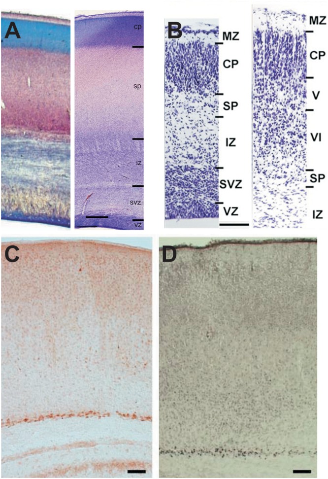
During early development the structure and function of the cerebral cortex is critically organized by subplate neurons (SPNs), a mostly transient population of glutamatergic and GABAergic neurons located below the cortical plate. At the molecular and morphological level SPNs represent a rather diverse population of cells expressing a variety of genetic markers and revealing different axonal-dendritic morphologies. Electrophysiologically SPNs are characterized by their rather mature intrinsic membrane properties and firing patterns. They are connected via electrical and chemical synapses to local and remote neurons, e.g., thalamic relay neurons forming the first thalamocortical input to the cerebral cortex. Therefore SPNs are robustly activated at pre- and perinatal stages by the sensory periphery. Although SPNs play pivotal roles in early neocortical activity, development and plasticity, they mostly disappear by programmed cell death during further maturation. On the one hand, SPNs may be selectively vulnerable to hypoxia-ischemia contributing to brain damage, on the other hand there is some evidence that enhanced survival rates or alterations in SPN distribution may contribute to the etiology of neurological or psychiatric disorders. This review aims to give a comprehensive and up-to-date overview on the many functions of SPNs during early physiological and pathophysiological development of the cerebral cortex.
Keywords: connectivity; development; neocortex; pathology; plasticity; review; subplate.
Figures

Similar articles
-
Changing subplate circuits: Early activity dependent circuit plasticity.Front Cell Neurosci. 2023 Jan 11;16:1067365. doi: 10.3389/fncel.2022.1067365. eCollection 2022. Front Cell Neurosci. 2023. PMID: 36713777 Free PMC article.
-
Transient Hypoxemia Chronically Disrupts Maturation of Preterm Fetal Ovine Subplate Neuron Arborization and Activity.J Neurosci. 2017 Dec 6;37(49):11912-11929. doi: 10.1523/JNEUROSCI.2396-17.2017. Epub 2017 Oct 31. J Neurosci. 2017. PMID: 29089437 Free PMC article.
-
The subplate and early cortical circuits.Annu Rev Neurosci. 2010;33:23-48. doi: 10.1146/annurev-neuro-060909-153244. Annu Rev Neurosci. 2010. PMID: 20201645 Review.
-
Subplate Neurons as an Organizer of Mammalian Neocortical Development.Front Neuroanat. 2020 Mar 19;14:8. doi: 10.3389/fnana.2020.00008. eCollection 2020. Front Neuroanat. 2020. PMID: 32265668 Free PMC article.
-
The Integrative Function of Silent Synapses on Subplate Neurons in Cortical Development and Dysfunction.Front Neuroanat. 2019 Apr 16;13:41. doi: 10.3389/fnana.2019.00041. eCollection 2019. Front Neuroanat. 2019. PMID: 31040772 Free PMC article. Review.
Cited by
-
Dynamic interplay between thalamic activity and Cajal-Retzius cells regulates the wiring of cortical layer 1.Cell Rep. 2022 Apr 12;39(2):110667. doi: 10.1016/j.celrep.2022.110667. Cell Rep. 2022. PMID: 35417707 Free PMC article.
-
A direct excitatory projection from entorhinal layer 6b neurons to the hippocampus contributes to spatial coding and memory.Nat Commun. 2022 Aug 16;13(1):4826. doi: 10.1038/s41467-022-32559-8. Nat Commun. 2022. PMID: 35974109 Free PMC article.
-
Changing subplate circuits: Early activity dependent circuit plasticity.Front Cell Neurosci. 2023 Jan 11;16:1067365. doi: 10.3389/fncel.2022.1067365. eCollection 2022. Front Cell Neurosci. 2023. PMID: 36713777 Free PMC article.
-
Emerging signs of autism spectrum disorder in infancy: Putative neural substrate.Dev Med Child Neurol. 2022 Nov;64(11):1344-1350. doi: 10.1111/dmcn.15333. Epub 2022 Jul 8. Dev Med Child Neurol. 2022. PMID: 35801808 Free PMC article. Review.
-
Altered synaptic connectivity in an in vitro human model of STXBP1 encephalopathy.Brain. 2023 Mar 1;146(3):850-857. doi: 10.1093/brain/awac396. Brain. 2023. PMID: 36315647 Free PMC article.
References
Publication types
LinkOut - more resources
Full Text Sources