

Are the Cognitive Alterations Present in Children Born From Preeclamptic Pregnancies the Result of Impaired Angiogenesis? Focus on the Potential Role of the VEGF Family
- PMID: 30487752
- PMCID: PMC6246680
- DOI: 10.3389/fphys.2018.01591
Are the Cognitive Alterations Present in Children Born From Preeclamptic Pregnancies the Result of Impaired Angiogenesis? Focus on the Potential Role of the VEGF Family
Abstract
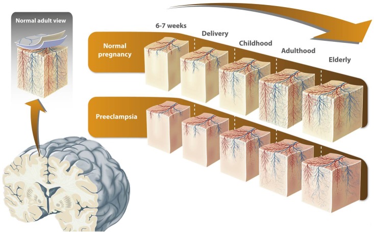
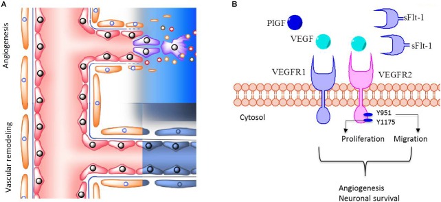
Evidence from clinical studies has proposed that children born from preeclamptic women have a higher risk of suffering neurological, psychological, or behavioral alterations. However, to date, the mechanisms behind these outcomes are poorly understood. Here, we speculate that the neurodevelopmental alterations in the children of preeclamptic pregnancies result from impaired angiogenesis. The pro-angiogenic factors vascular endothelial growth factor (VEGF) and placental growth factor (PlGF) are key regulators of both vascular and neurological development, and it has been widely demonstrated that umbilical blood of preeclamptic pregnancies contains high levels of soluble VEGF receptor type 1 (sFlt-1), a decoy receptor of VEGF. As a consequence, this anti-angiogenic state could lead to long-lasting neurological outcomes. In this non-systematic review, we propose that alterations in the circulating concentrations of VEGF, PlGF, and sFlt-1 in preeclamptic pregnancies will affect both fetal cerebrovascular function and neurodevelopment, which in turn may cause cognitive alterations in post-natal life.
Keywords: angiogenesis; neurocognitive; neurovascular; placental growth factor; preeclampsia; sFlt-1; vascular endothelial growth factor.
Figures
Similar articles
-
Altered angiogenesis in preeclampsia: evaluation of a new test system for measuring placental growth factor.Clin Chem Lab Med. 2007;45(11):1504-10. doi: 10.1515/CCLM.2007.316. Clin Chem Lab Med. 2007. PMID: 17970705
-
Dysregulation of circulating autoantibodies against VEGF-A, VEGFR-1 and PlGF in preeclampsia - A role in placental and vascular health?Pregnancy Hypertens. 2017 Oct;10:83-89. doi: 10.1016/j.preghy.2017.06.002. Epub 2017 Jun 27. Pregnancy Hypertens. 2017. PMID: 29153696
-
In vivo uteroplacental release of placental growth factor and soluble Fms-like tyrosine kinase-1 in normal and preeclamptic pregnancies.Am J Obstet Gynecol. 2016 Dec;215(6):782.e1-782.e9. doi: 10.1016/j.ajog.2016.07.056. Epub 2016 Aug 5. Am J Obstet Gynecol. 2016. PMID: 27503620
-
Pro-angiogenic therapeutics for preeclampsia.Biol Sex Differ. 2018 Aug 25;9(1):36. doi: 10.1186/s13293-018-0195-5. Biol Sex Differ. 2018. PMID: 30144822 Free PMC article. Review.
-
Impaired adenosine-mediated angiogenesis in preeclampsia: potential implications for fetal programming.Front Pharmacol. 2014 Jun 5;5:134. doi: 10.3389/fphar.2014.00134. eCollection 2014. Front Pharmacol. 2014. PMID: 24926270 Free PMC article. Review.
Cited by
-
Development of the Neuro-Immune-Vascular Plexus in the Ventricular Zone of the Prenatal Rat Neocortex.Cereb Cortex. 2021 Mar 5;31(4):2139-2155. doi: 10.1093/cercor/bhaa351. Cereb Cortex. 2021. PMID: 33279961 Free PMC article.
-
Systemic Maternal Human sFLT1 Overexpression Leads to an Impaired Foetal Brain Development of Growth-Restricted Foetuses upon Experimental Preeclampsia.Oxid Med Cell Longev. 2022 Jun 2;2022:3024032. doi: 10.1155/2022/3024032. eCollection 2022. Oxid Med Cell Longev. 2022. PMID: 35693702 Free PMC article.
-
Association of Preeclampsia in Term Births With Neurodevelopmental Disorders in Offspring.JAMA Psychiatry. 2020 Aug 1;77(8):823-829. doi: 10.1001/jamapsychiatry.2020.0306. JAMA Psychiatry. 2020. PMID: 32236510 Free PMC article.
-
Antioxidants Attenuate Heat Shock Induced Premature Senescence of Bovine Mesenchymal Stem Cells.Int J Mol Sci. 2022 May 20;23(10):5750. doi: 10.3390/ijms23105750. Int J Mol Sci. 2022. PMID: 35628565 Free PMC article.
-
Early Abnormal Placentation and Evidence of Vascular Endothelial Growth Factor System Dysregulation at the Feto-Maternal Interface After Periconceptional Alcohol Consumption.Front Physiol. 2022 Feb 2;12:815760. doi: 10.3389/fphys.2021.815760. eCollection 2021. Front Physiol. 2022. PMID: 35185604 Free PMC article. Review.
References
-
- ACOG (2013). Hypertension in Pregnancy. Washington, DC: ACOG.
-
- Acurio J., Herlitz K., Troncoso F., Aguayo C., Bertoglia P., Escudero C. (2016). Adenosine A2A receptor regulates expression of vascular endothelial growth factor in feto-placental endothelium from normal and late-onset pre-eclamptic pregnancies. Purinergic Signal. 13 51–60. 10.1007/s11302-016-9538-z - DOI - PMC - PubMed
LinkOut - more resources
Full Text Sources
Research Materials