

Silencing of Hypoglossal Motoneurons Leads to Sleep Disordered Breathing in Lean Mice
- PMID: 30487776
- PMCID: PMC6246694
- DOI: 10.3389/fneur.2018.00962
Silencing of Hypoglossal Motoneurons Leads to Sleep Disordered Breathing in Lean Mice
Abstract
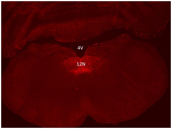
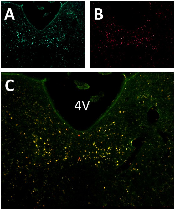
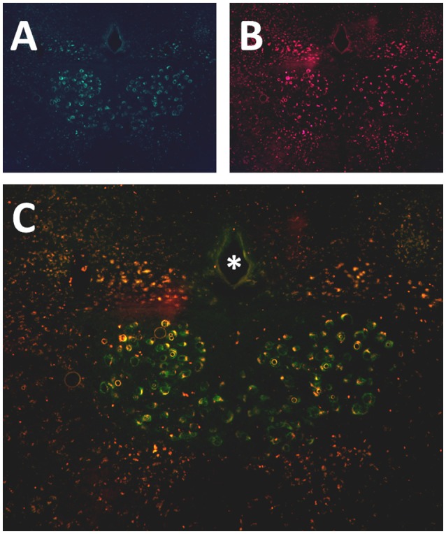
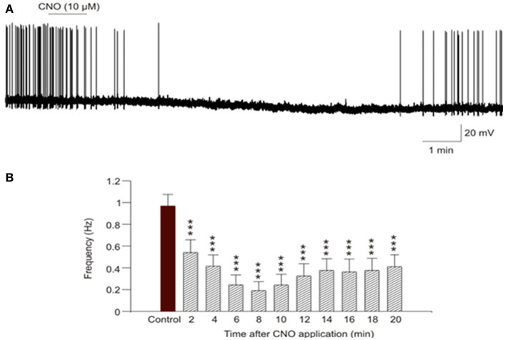
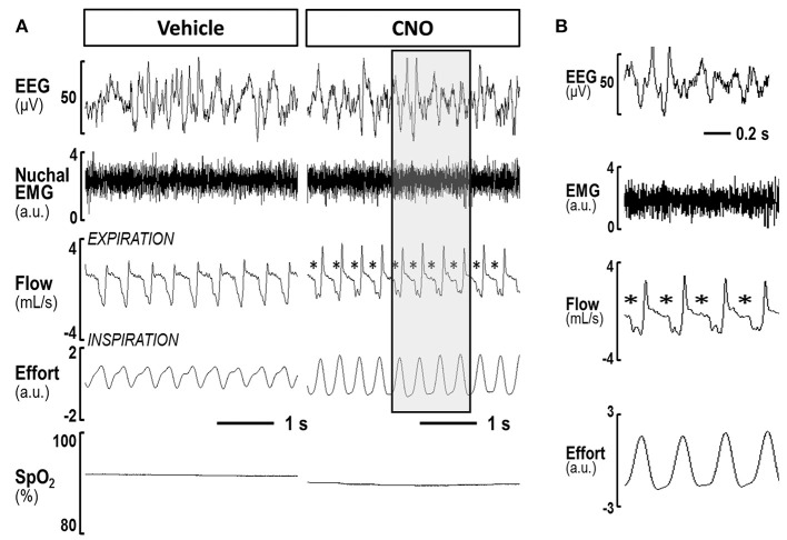
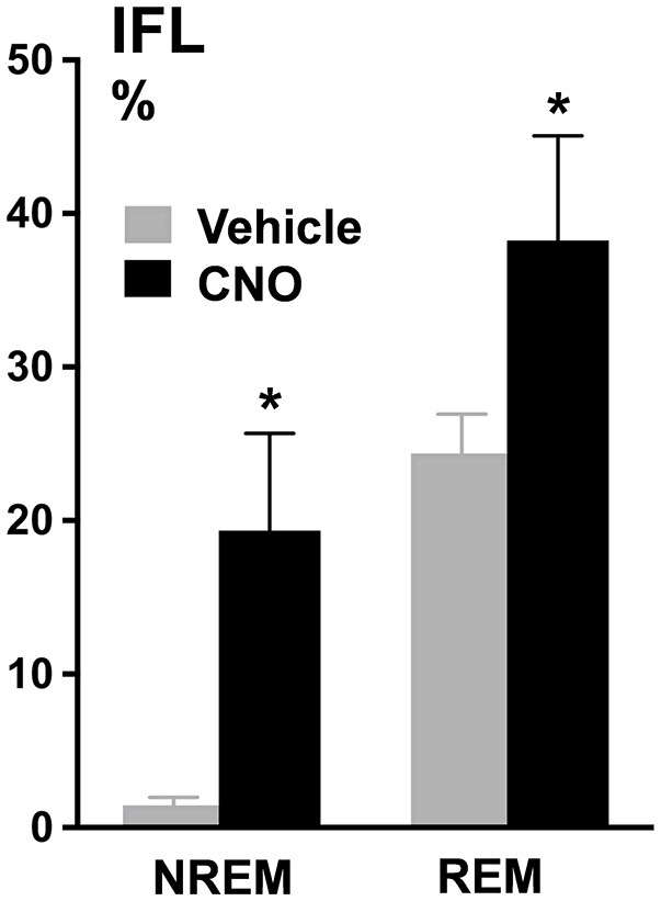
Obstructive Sleep Apnea (OSA) is a prevalent condition and a major cause of morbidity and mortality in Western Society. The loss of motor input to the tongue and specifically to the genioglossus muscle during sleep is associated with pharyngeal collapsibility and the development of OSA. We applied a novel chemogenetic method to develop a mouse model of sleep disordered breathing Our goal was to reversibly silence neuromotor input to the genioglossal muscle using an adeno-associated viral vector carrying inhibitory designer receptors exclusively activated by designer drugs AAV5-hM4Di-mCherry (DREADD), which was delivered bilaterally to the hypoglossal nucleus in fifteen C57BL/6J mice. In the in vivo experiment, 4 weeks after the viral administration mice were injected with a DREADD ligand clozapine-N-oxide (CNO, i.p., 1mg/kg) or saline followed by a sleep study; a week later treatments were alternated and a second sleep study was performed. Inspiratory flow limitation was recognized by the presence of a plateau in mid-respiratory flow; oxyhemoglobin desaturations were defined as desaturations >4% from baseline. In the in vitro electrophysiology experiment, four males and three females of 5 days of age were used. Sixteen-nineteen days after DREADD injection brain slices of medulla were prepared and individual hypoglossal motoneurons were recorded before and after CNO application. Positive mCherry staining was detected in the hypoglossal nucleus in all mice confirming successful targeting. In sleep studies, CNO markedly increased the frequency of flow limitation n NREM sleep (from 1.9 ± 1.3% after vehicle injection to 14.2 ± 3.4% after CNO, p < 0.05) and REM sleep (from 22.3% ± 4.1% to 30.9 ± 4.6%, respectively, p < 0.05) compared to saline treatment, but there was no significant oxyhemoglobin desaturation or sleep fragmentation. Electrophysiology recording in brain slices showed that CNO inhibited firing frequency of DREADD-containing hypoglossal motoneurons. We conclude that chemogenetic approach allows to silence hypoglossal motoneurons in mice, which leads to sleep disordered breathing manifested by inspiratory flow limitation during NREM and REM sleep without oxyhemoglobin desaturation or sleep fragmentation. Other co-morbid factors, such as compromised upper airway anatomy, may be needed to achieve recurrent pharyngeal obstruction observed in OSA.
Keywords: chemogenetic; neuromuscular activity; obstructive sleeep apnea; sleep; upper airway.
Figures
Similar articles
-
Chemogenetic activation of hypoglossal motoneurons in a mouse model of Pompe disease.J Neurophysiol. 2022 Nov 1;128(5):1133-1142. doi: 10.1152/jn.00026.2022. Epub 2022 Aug 17. J Neurophysiol. 2022. PMID: 35976060 Free PMC article.
-
Designer Receptors Exclusively Activated by Designer Drugs Approach to Treatment of Sleep-disordered Breathing.Am J Respir Crit Care Med. 2021 Jan 1;203(1):102-110. doi: 10.1164/rccm.202002-0321OC. Am J Respir Crit Care Med. 2021. PMID: 32673075 Free PMC article.
-
Chemogenetic stimulation of the hypoglossal neurons improves upper airway patency.Sci Rep. 2017 Mar 10;7:44392. doi: 10.1038/srep44392. Sci Rep. 2017. PMID: 28281681 Free PMC article.
-
Respiratory motor activity: influence of neuromodulators and implications for sleep disordered breathing.Can J Physiol Pharmacol. 2007 Jan;85(1):155-65. doi: 10.1139/y06-089. Can J Physiol Pharmacol. 2007. PMID: 17487255 Review.
-
Neuroanatomical Basis of State-Dependent Activity of Upper Airway Muscles.Front Neurol. 2018 Sep 10;9:752. doi: 10.3389/fneur.2018.00752. eCollection 2018. Front Neurol. 2018. PMID: 30250449 Free PMC article. Review.
Cited by
-
Chemogenetic activation of hypoglossal motoneurons in a mouse model of Pompe disease.J Neurophysiol. 2022 Nov 1;128(5):1133-1142. doi: 10.1152/jn.00026.2022. Epub 2022 Aug 17. J Neurophysiol. 2022. PMID: 35976060 Free PMC article.
-
Detection of central and obstructive sleep apneas in mice: A new surgical and recording protocol.PLoS One. 2025 Mar 28;20(3):e0320650. doi: 10.1371/journal.pone.0320650. eCollection 2025. PLoS One. 2025. PMID: 40153464 Free PMC article.
-
Management of Obstructive Sleep Apnea in Patients With Heart Failure.Front Med (Lausanne). 2022 Feb 18;9:803388. doi: 10.3389/fmed.2022.803388. eCollection 2022. Front Med (Lausanne). 2022. PMID: 35252246 Free PMC article. Review.
-
Multilevel Airway Obstruction Phenotypes in Adult OSA.OTO Open. 2023 Jun 23;7(2):e21. doi: 10.1002/oto2.21. eCollection 2023 Apr-Jun. OTO Open. 2023. PMID: 37359981 Free PMC article.
-
Intranasal Leptin Prevents Opioid-induced Sleep-disordered Breathing in Obese Mice.Am J Respir Cell Mol Biol. 2020 Oct;63(4):502-509. doi: 10.1165/rcmb.2020-0117OC. Am J Respir Cell Mol Biol. 2020. PMID: 32603263 Free PMC article.
References
Grants and funding
LinkOut - more resources
Full Text Sources
Other Literature Sources