

Schistosome Eggs Impair Protective Th1/Th17 Immune Responses Against Salmonella Infection
- PMID: 30487793
- PMCID: PMC6246638
- DOI: 10.3389/fimmu.2018.02614
Schistosome Eggs Impair Protective Th1/Th17 Immune Responses Against Salmonella Infection
Abstract
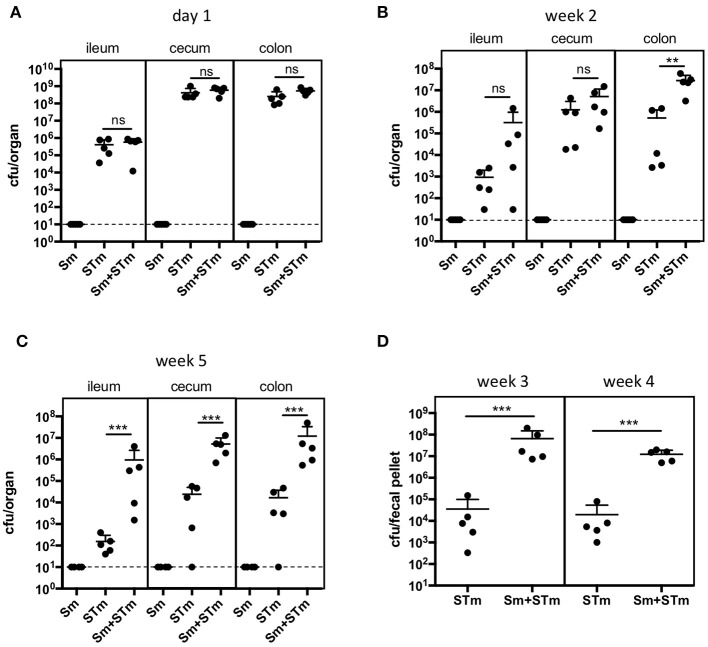
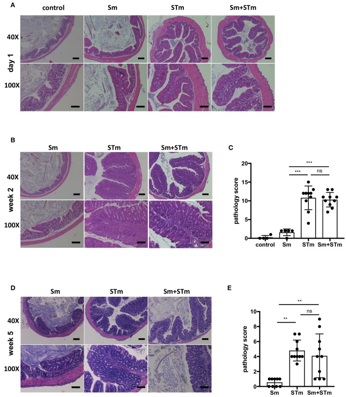
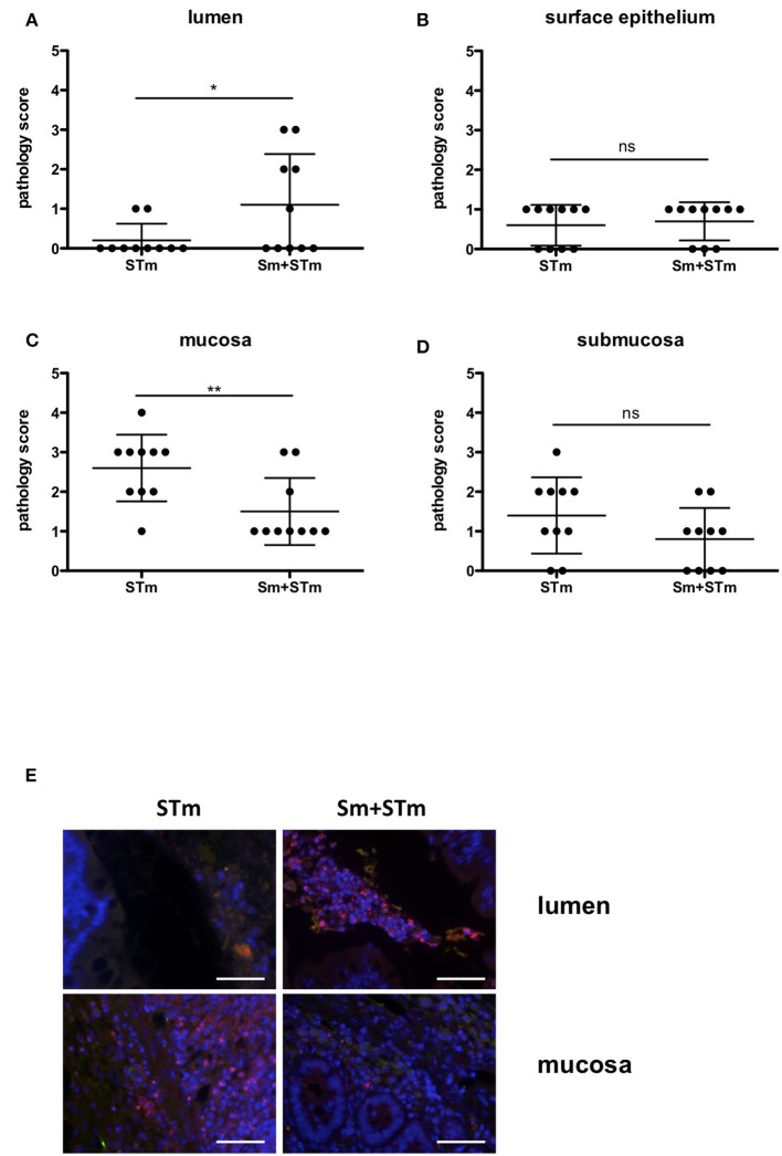
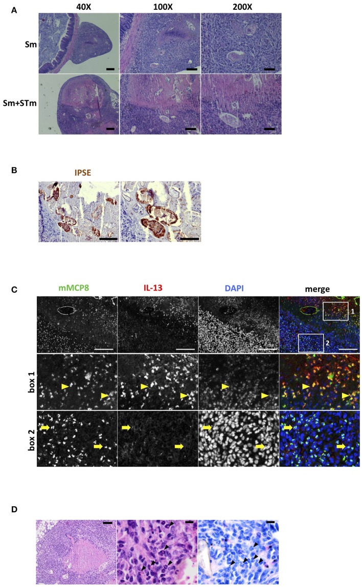
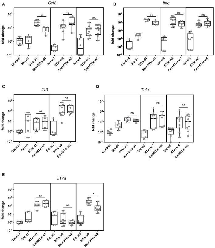
Countries with a high incidence of helminth infections are characterized by high morbidity and mortality to infections with intracellular pathogens such as Salmonella. Some patients with Salmonella-Schistosoma co-infections develop a so-called "chronic septicemic salmonellosis," with prolonged fever and enlargement of the liver and spleen. These effects are most likely due to the overall immunoregulatory activities of schistosomes such as induction of Tregs, Bregs, alternatively activated macrophages, and degradation of antibodies. However, detailed underlying mechanisms are not very well investigated. Here, we show that intraperitoneal application of live Schistosoma mansoni eggs prior to infection with Salmonella Typhimurium in mice leads to an impairment of IFN-γ and IL-17 responses together with a higher bacterial load compared to Salmonella infection alone. S. mansoni eggs were found in granulomas in the visceral peritoneum attached to the colon. Immunohistological staining revealed IPSE/alpha-1, a glycoprotein secreted from live schistosome eggs, and recruited basophils around the eggs. Noteworthy, IPSE/alpha-1 is known to trigger IL-4 and IL-13 release from basophils which in turn is known to suppress Th1/Th17 responses. Therefore, our data support a mechanism of how schistosomes impair a protective immune response against Salmonella infection and increase our understanding of helminth-bacterial co-infections.
Keywords: IPSE/alpha-1; Salmonella Typhimurium; Schistosoma mansoni; Th1; Th17; co-infection; immunoregulation.
Figures
Similar articles
-
Schistosoma mansoni Egg-Released IPSE/alpha-1 Dampens Inflammatory Cytokine Responses via Basophil Interleukin (IL)-4 and IL-13.Front Immunol. 2018 Oct 10;9:2293. doi: 10.3389/fimmu.2018.02293. eCollection 2018. Front Immunol. 2018. PMID: 30364177 Free PMC article.
-
IPSE/alpha-1 of Schistosoma mansoni egg induces enlargement of granuloma but does not alter Th2 balance after infection.Parasite Immunol. 2010 May;32(5):345-53. doi: 10.1111/j.1365-3024.2009.01192.x. Parasite Immunol. 2010. PMID: 20500663
-
Strongyloides venezuelensis-infection alters the profile of cytokines and liver inflammation in mice co-infected with Schistosoma mansoni.Cytokine. 2020 Mar;127:154931. doi: 10.1016/j.cyto.2019.154931. Epub 2019 Nov 26. Cytokine. 2020. PMID: 31783260
-
Human schistosomiasis mansoni: immune responses during acute and chronic phases of the infection.Acta Trop. 2008 Nov-Dec;108(2-3):109-17. doi: 10.1016/j.actatropica.2008.05.027. Epub 2008 Jun 5. Acta Trop. 2008. PMID: 18577364 Review.
-
Infection by Schistosoma mansoni during pregnancy: Effects on offspring immunity.Life Sci. 2017 Sep 15;185:46-52. doi: 10.1016/j.lfs.2017.07.021. Epub 2017 Jul 25. Life Sci. 2017. PMID: 28754617 Review.
Cited by
-
Blood Flukes and Arterial Damage: A Review of Aneurysm Cases in Patients with Schistosomiasis.Can J Infect Dis Med Microbiol. 2022 Dec 3;2022:6483819. doi: 10.1155/2022/6483819. eCollection 2022. Can J Infect Dis Med Microbiol. 2022. PMID: 36510604 Free PMC article. Review.
-
Infection with soil-transmitted helminths and their impact on coinfections.Front Parasitol. 2023 May 24;2:1197956. doi: 10.3389/fpara.2023.1197956. eCollection 2023. Front Parasitol. 2023. PMID: 39816832 Free PMC article. Review.
-
Schistosomiasis-Microbiota Interactions: A Systematic Review and Meta-Analysis.Pathogens. 2024 Oct 16;13(10):906. doi: 10.3390/pathogens13100906. Pathogens. 2024. PMID: 39452777 Free PMC article.
-
Schistosoma mansoni Eggs Modulate the Timing of Granuloma Formation to Promote Transmission.Cell Host Microbe. 2021 Jan 13;29(1):58-67.e5. doi: 10.1016/j.chom.2020.10.002. Epub 2020 Oct 28. Cell Host Microbe. 2021. PMID: 33120115 Free PMC article.
-
Prevalence and Clinical Relevance of Schistosoma mansoni Co-Infection with Mycobacterium tuberculosis: A Systematic Literature Review.Open J Epidemiol. 2023 Feb;13(1):97-111. doi: 10.4236/ojepi.2023.131008. Epub 2023 Feb 23. Open J Epidemiol. 2023. PMID: 36910425 Free PMC article.
References
-
- Gendrel D, Kombila M, Beaudoin-Leblevec G, Richard-Lenoble D. Nontyphoidal salmonellal septicemia in Gabonese children infected with Schistosoma intercalatum. Clin Infect Dis. (1994) 18:103–5. - PubMed
Publication types
MeSH terms
Substances
LinkOut - more resources
Full Text Sources
Medical
Molecular Biology Databases