. 2018 Nov 14:9:2645.
doi: 10.3389/fimmu.2018.02645. eCollection 2018.

Site-Specific N-Glycan Characterization of Grass Carp Serum IgM

Affiliations

Site-Specific N-Glycan Characterization of Grass Carp Serum IgM

Yi-Ling Su et al. Front Immunol. .

Abstract

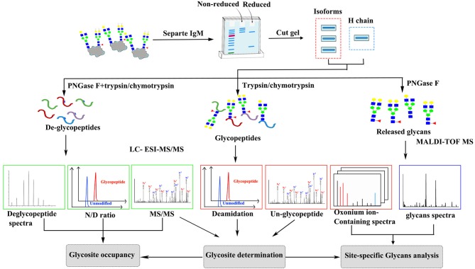
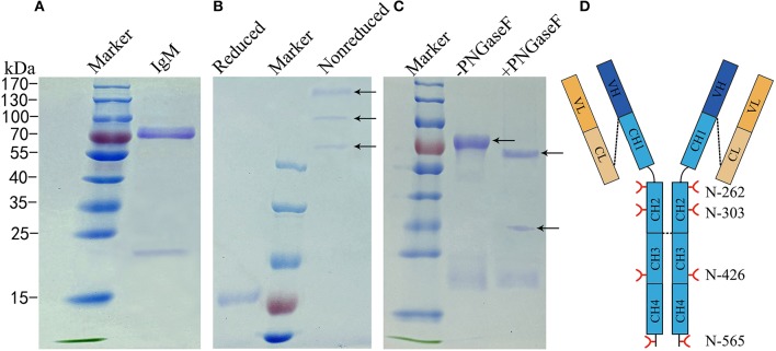
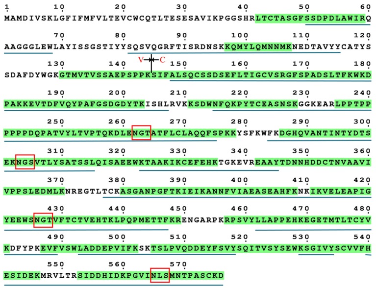
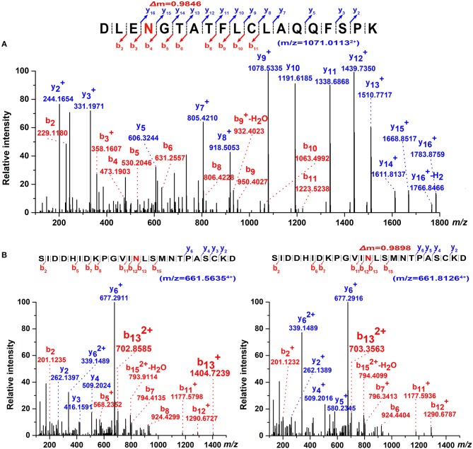
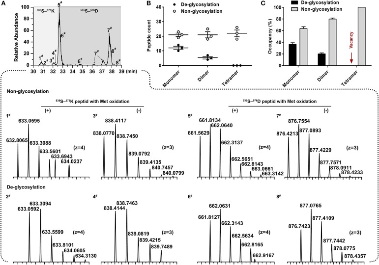
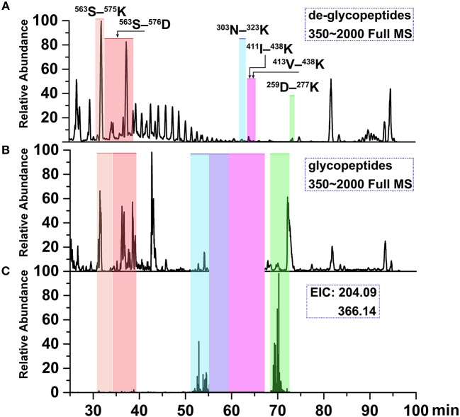
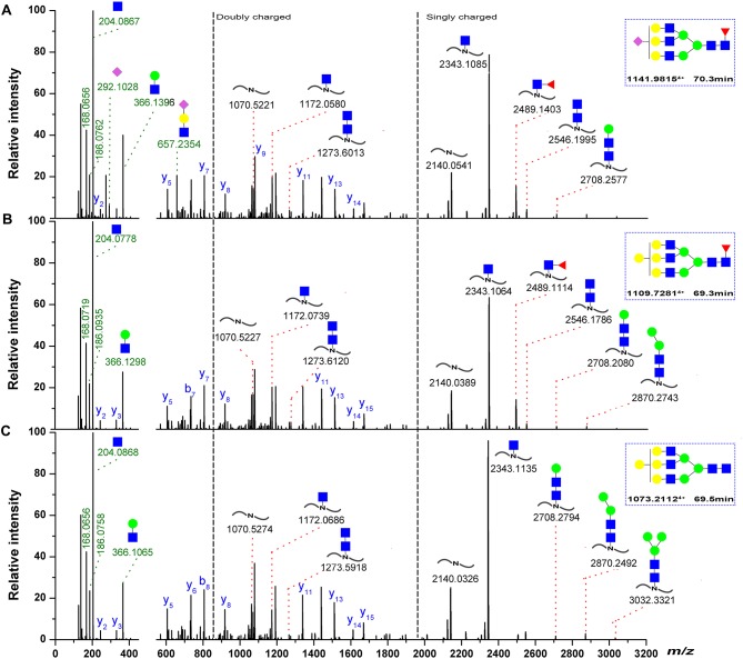
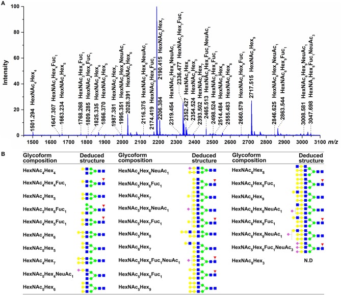
Immunoglobulin M (IgM) is the major antibody in teleost fish and plays an important role in humoral adaptive immunity. The N-linked carbohydrates presenting on IgM have been well documented in higher vertebrates, but little is known regarding site-specific N-glycan characteristics in teleost IgM. In order to characterize these site-specific N-glycans, we conducted the first study of the N-glycans of each glycosylation site of the grass carp serum IgM. Among the four glycosylation sites, the Asn-262, Asn-303, and Asn-426 residues were efficiently glycosylated, while Asn-565 at the C-terminal tailpiece was incompletely occupied. A striking decrease in the level of occupancy at the Asn-565 glycosite was observed in dimeric IgM compared to that in monomeric IgM, and no glycan occupancy of Asn-565 was observed in tetrameric IgM. Glycopeptide analysis with liquid chromatography-electrospray ionization tandem mass spectrometry revealed mainly complex-type glycans with substantial heterogeneity, with neutral; monosialyl-, disialyl- and trisialylated; and fucosyl-and non-fucosyl-oligosaccharides conjugated to grass carp serum IgM. Glycan variation at a single site was greatest at the Asn-262 glycosite. Unlike IgMs in other species, only traces of complex-type and no high-mannose glycans were found at the Asn-565 glycosite. Matrix-assisted laser desorption ionization analysis of released glycans confirmed the overwhelming majority of carbohydrates were of the complex-type. These results indicate that grass carp serum IgM exhibits unique N-glycan features and highly processed oligosaccharides attached to individual glycosites.

Keywords: N-glycan; glycosylation; grass carp; immunoglobulin M; liquid chromatography-electrospray ionization tandem mass spectrometry (LC-ESI-MS/MS); matrix assisted laser desorption/ionization-time-of-flight-MS (MALDI-TOF-MS); teleost.


Figures

Figure 1
Figure 1
Scheme of the experimental approach to characterize grass carp serum IgM glycoform.
Figure 2
Figure 2
Analysis of N-glycans assignments on grass carp serum IgM. (A) A 4–10% uncontinued SDS-PAGE of reduced samples to show 75–80 kDa heavy chain band and 23 kDa light chain band of grass carp IgM. (B) A 6% SDS-PAGE stained Coomassie Blue with showing different grass carp IgM isoforms, in the non-reduced samples lane, from up to down, the tetramer, dimer, and monomer were marked by arrow. (C) IgM digested with (+) or without (–) PNGase F, arrow at the bottom indicate PNGase F. (D) Schematic of four N-glycosyationsites on grass carp IgM, red forks represent four putative N-glycosylation sites on heavy chain, black lines at the end of CH4 domain indicating 16–18 amino acid residue at the C-terminal tailpiece, possible inter-subunit disulfide bridges between heavy chains and covalently link the light chain are shown with black dash lines.
Figure 3
Figure 3
Sequence coverage of grass carp IgM heavy chain. The identified tryptic peptides are indicated by green shadow, four putative N-glycosylation sites are marked by red box. The identified chymotryptic peptides are indicated by black underline.
Figure 4
Figure 4
Determination of N-glycosylation sites by high-resolution ESI MS. (A) Representative MS/MS spectrum of the tryptic 259D–277Kpeptide (m/z = 1071.01132+) covering Asn-262 glycosite with PNGase F digestion. The mass increment of 0.9846 Da comparing to its theoretical mass weight is indicated. (B) Dimer-isotopic mass spectrum of 553S–576D peptide covering Asn-565 glycosite. Non-glycosylated 553S–576D peptide (m/z = 661.56354+) is shown in left panel, while de-glycosylated 553S–576D peptide (m/z = 661.81264+) with a mass increasing of 0.9898 Da due to the converting of Asn-565 residue to an Asp with PNGase F treatment is indicated in right panel.
Figure 5
Figure 5
Determination of site occupancy for Asn-565 glycosite. (A) Overlaid chromatograms of non-glycosylated and de-glycosylated Asn-565 peptides. A total of 8 peptide variants were identified; 1# and 3#, 5# and 7#, non-glycosylated 553S–575K peptide and 553S–576D peptide without, and with methionineoxidation. 2# and 4#, 6# and 8#, De-glycosylated 553S–575K peptide and 553S–576D peptide without, and with methionineoxidation. (B) Asn-565 peptides spectral counts plot. Non-glycosylated or de-glycosylated Asn-565 peptides counts are shown by the sum of 553S–575K peptide and 553S–576D peptide. (C) Asn-565 glycosite occupancy. Occupancy was calculated by sum of the non-glycosylated or de-glycosylated peptide as a percentage of the total spectral counts of non-glycosylated and de-glycosylated peptide. Error bars in (B,C) indicate the standard deviation of the same serum pools with three repeats.
Figure 6
Figure 6
Ion chromatograms of LC-separated tryptic peptides. (A) Base peak chromatogram (BPC) of the tryptic de-glycopeptides with PNGase F digestion. (B) BPC of tryptic glycopeptides with HILIC enrichment. (C) Extracted ion chromatograms (EICs) of diagnostic oxonium fragment ions at m/z of 204.09 and 366.14. The shaded regions depict the identified deglycopeptides, glycopeptide clusters and Tables 2, 3 summarizes glycan heterogeneity at each site.
Figure 7
Figure 7
Representative spectra for Asn-262 glycopeptides with a sialylated glycan. (A) A neutral glycan with core-fucose (B) A neutral glycan without core-fucose glycan. (C) The oxonium ions were used to extract the intact glycopeptide spectrum, and the masses of the precursor and peptide/peptide + core GlcNAc ± Fuc ± Hex ions (red) can provide structural details, the b/y-ions (only y-ions were marked, blue) of the peptide portion were used to confirm peptide sequence. The proposed glycan structures were determined on the basis of monosaccharide composition, the monosaccharide linkages were not yet determined. Symbols as the following: blue box, N-acetylglucosamine; green circle, mannose; yellow circle, galactose; red triangle, fucose; pink diamond, N-acetylneuraminic acid.
Figure 8
Figure 8
Glycans detected by MALDI-TOF. (A) Molecular masses are of unlabeled glycans, detected as [M + Na]+ in positive polarity mode. (B) The proposed glycan structures were determined on the basis of monosaccharide composition, as well as literature (24, 25, 48). For symbol definitions, seen Figure 6. N.D., not determined.

Similar articles

Cited by

References

    1. Partridge EA, Le Roy C, Di Guglielmo GM, Pawling J, Cheung P, Granovsky M, et al. . Regulation of cytokine receptors by Golgi N-glycan processing and endocytosis. Science (2004) 306:120–4. 10.1126/science.1102109 - DOI - PubMed
    1. Dennis JW, Nabi IR, Demetriou M. Metabolism, cell surface organization, and disease. Cell (2009) 139:1229–41. 10.1016/j.cell.2009.12.008 - DOI - PMC - PubMed
    1. Starossom SC, Mascanfroni ID, Imitola J, Cao L, Raddassi K, Hernandez SF, et al. . Galectin-1 deactivates classically activated microglia and protects from inflammation-induced neurodegeneration. Immunity (2012) 37:249–63. 10.1016/j.immuni.2012.05.023 - DOI - PMC - PubMed
    1. Vasta GR, Ahmed H, Nita-Lazar M, Banerjee A, Pasek M, Shridhar S, et al. . Galectins as self/non-self recognition receptors in innate and adaptive immunity: an unresolved paradox. Front Immunol. (2012) 3:199. 10.3389/fimmu.2012.00199 - DOI - PMC - PubMed
    1. Rudd PM, Elliott T, Cresswell P, Wilson IA, Dwek RA. Glycosylation and the immune system. Science (2001) 291:2370–6. 10.1126/science.291.5512.2370 - DOI - PubMed

Publication types