

Neuroimaging in cockroach phobia: An experimental study
- PMID: 30487896
- PMCID: PMC6220927
- DOI: 10.1016/j.ijchp.2017.06.002
Neuroimaging in cockroach phobia: An experimental study
Abstract
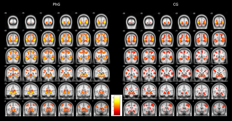
In this study we explored the neuroimaging characteristics of persons with specific small animal (cockroach) phobia to determine whether there are differences in cerebral activity between persons with and without cockroach phobia under conditions of phobic and non-phobic stimulation. Method: 24 adult persons (12 with phobia) were studied. The diagnosis of phobia was obtained with a structured interview and questionnaires. All participants were exposed to a 3D video presentation during an fMRI session. Results: The phobic group showed significant differential activations that were congruent with a dual route model of fear processing through the thalamus-amygdala (route I) and the thalamus-sensory and association cortex-entorhinal cortex-hippocampus-subiculum-amygdala (route II). Apart from this dual route, we also found differential activations in the globus pallidum, parahippocampal gyrus, insula, pars orbitalis, triangularis and opercularis of the frontal cortex, and cerebellum. Respect to non-phobic group, no activations were found in the insula or the anterior cingulate cortex. Conclusions: There seems to be a dual route depending on how persons with phobia to cockroaches process phobic stimuli. This double processing can have implications for the psychological treatment of specific phobias.
Antecedentes/objetivo: En este estudio se exploran las características en neuroimagen de personas con fobia específica a pequeños animales (cucarachas), para determinar si existen diferencias en la actividad cerebral entre personas con y sin fobia a las cucarachas, bajo condiciones de estimulación fóbica y no fóbica. Método: Se estudiaron 24 adultos (12 con fobia). El diagnóstico de fobia específica se obtuvo mediante una entrevista estructurada y cuestionarios. Todos fueron expuestos a una presentación en video 3D durante una sesión de RMNf.
Resultados: El grupo con fobia mostró activaciones diferenciales significativas, que fueron congruentes con el modelo de doble ruta en el procesamiento del miedo, a través del tálamo-amígdala (ruta I), y tálamo-corteza entorrinal-hipocampo-subículo-amígdala (ruta II). Además, se encontraron activaciones diferenciales en el globo pálido, en el giro hipocampal, ínsula, y en los pars orbitalis, triangularis y opercularis. Con respecto al grupo control, no se observaron activaciones de la ínsula ni el cingulado.
Conclusiones: Parece evidenciarse un modelo de doble ruta en el procesamiento de estímulos fóbicos. Este doble proceso puede tener implicaciones para el tratamiento psicológico de las fobias específicas.
Keywords: Cockroaches; Experimental study; Neuroimage; Specific phobia; fMRI.
Figures
Similar articles
-
Weaving the (neuronal) web: fear learning in spider phobia.Neuroimage. 2011 Jan 1;54(1):681-8. doi: 10.1016/j.neuroimage.2010.07.049. Epub 2010 Jul 27. Neuroimage. 2011. PMID: 20673801
-
Networks of phobic fear: Functional connectivity shifts in two subtypes of specific phobia.Neurosci Lett. 2018 Jan 1;662:167-172. doi: 10.1016/j.neulet.2017.10.031. Epub 2017 Oct 18. Neurosci Lett. 2018. PMID: 29054435
-
The functional neuroanatomy of blood-injection-injury phobia: a comparison with spider phobics and healthy controls.Psychol Med. 2010 Jan;40(1):125-34. doi: 10.1017/S0033291709005972. Epub 2009 May 13. Psychol Med. 2010. PMID: 19435544
-
Meta-analysis of functional brain imaging in specific phobia.Psychiatry Clin Neurosci. 2013 Jul;67(5):311-22. doi: 10.1111/pcn.12055. Epub 2013 May 28. Psychiatry Clin Neurosci. 2013. PMID: 23711114 Review.
-
Functional neuroimaging in specific phobia.Psychiatry Res. 2012 Jun 30;202(3):181-97. doi: 10.1016/j.pscychresns.2011.10.009. Epub 2012 Jul 16. Psychiatry Res. 2012. PMID: 22804970 Review.
Cited by
-
Neuronal Activity during Exposure to Specific Phobia through fMRI: Comparing Therapeutic Components of Cognitive Behavioral Therapy.Life (Basel). 2022 Jul 27;12(8):1132. doi: 10.3390/life12081132. Life (Basel). 2022. PMID: 36013311 Free PMC article.
-
Neuronal Correlates of Small Animal Phobia in Human Subjects through fMRI: The Role of the Number and Proximity of Stimuli.Life (Basel). 2021 Mar 26;11(4):275. doi: 10.3390/life11040275. Life (Basel). 2021. PMID: 33810230 Free PMC article.
-
Changes in Brain Activation through Cognitive-Behavioral Therapy with Exposure to Virtual Reality: A Neuroimaging Study of Specific Phobia.J Clin Med. 2021 Aug 9;10(16):3505. doi: 10.3390/jcm10163505. J Clin Med. 2021. PMID: 34441804 Free PMC article.
-
ENIGMA-anxiety working group: Rationale for and organization of large-scale neuroimaging studies of anxiety disorders.Hum Brain Mapp. 2022 Jan;43(1):83-112. doi: 10.1002/hbm.25100. Epub 2020 Jul 3. Hum Brain Mapp. 2022. PMID: 32618421 Free PMC article. Review.
-
Cockroaches are scarier than snakes and spiders: Validation of an affective standardized set of animal images (ASSAI).Behav Res Methods. 2021 Dec;53(6):2338-2350. doi: 10.3758/s13428-021-01577-7. Epub 2021 Apr 7. Behav Res Methods. 2021. PMID: 33826093 Free PMC article.
References
-
- Agustín-Pavón C. El miedo en el cerebro. Mente y Cerebro. 2013;58:40–41.
-
- Agustín-Pavón C., Braesicke K., Shiba Y., Santangelo A.M., Mikheenko Y., Cockroft G., Asma F., Clarke H., Man M.S., Roberts A.C. Lesions of Ventrolateral Prefrontal or Anterior Orbitofrontal Cortex in Primates Heighten Negative Emotion. Biological Psychiatry. 2012;72:266–272. - PubMed
-
- American Psychiatry Association, APA . Fifth Edition. Author; Washington, DC: 2013. Diagnostic and Statistical Manual of Mental Disorders.
-
- Apps R., Strata P. Neuronal circuits for fear and anxiety - the missing link. Nature Reviews Neuroscience. 2015;16:642–643. - PubMed
LinkOut - more resources
Full Text Sources
Miscellaneous
