

Transtentorial herniation from tumefactive multiple sclerosis mimicking primary brain tumor
- PMID: 30488006
- PMCID: PMC6213805
- DOI: 10.4103/sni.sni_131_18
Transtentorial herniation from tumefactive multiple sclerosis mimicking primary brain tumor
Abstract
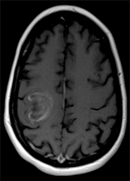
Background: Multiple sclerosis (MS) is a chronic central nervous system inflammatory demyelinating disease characterized by multiple lesions disseminated in time and space. The lesions often have characteristic imaging findings on magnetic resonance (MR) imaging and cerebrospinal fluid findings that lead to their diagnosis. At times, these lesions may resemble tumors due to their large size (>2 cm), significant vasogenic edema, and ring-enhancing MR imaging findings. Such lesions are described as tumefactive demyelinating lesions or tumefactive MS, and they are generally seen in aggressive forms of MS associated with rapid progression.
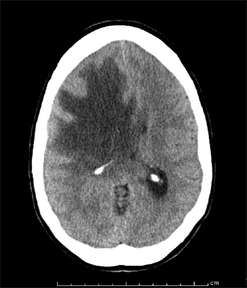
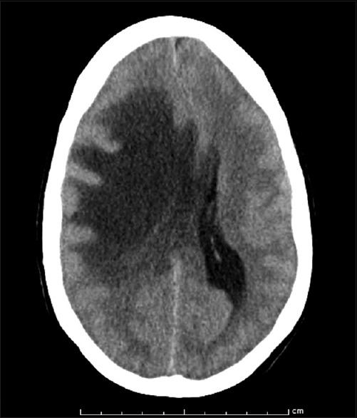
Case description: We report an uncommon but clinically significant case of transtentorial brain herniation secondary to malignant cerebral edema from tumefactive MS in a 50-year-old woman. After the initial diagnosis of MS, the patient continued to have progression of her white matter lesions suggesting evolution of her MS despite treatment with intravenous (IV) steroids, IV immunoglobulin, and plasmapheresis. She was admitted to the hospital with a new, large, ring-enhancing lesion that displayed significant mass effect from vasogenic edema and progressed, necessitating a decompressive hemicraniectomy.
Conclusion: Tumefactive MS presents a unique pathology that can often mimic primary brain tumors. Although these lesions affect white matter and infrequently cause a significant amount of mass effect, they can act like a tumor, causing edema that generates sufficient intracranial pressure to cause transtentorial herniation.
Keywords: Decompressive hemicraniectomy; transtentorial herniation; tumefactive demyelinating lesion; tumefactive multiple sclerosis.
Conflict of interest statement
There are no conflicts of interest.
Figures
Similar articles
-
Tumefactive Multiple Sclerosis, A Rare Variant Presenting as Multiple Ring-enhancing Lesions in an Immunocompetent Patient: A Case Report.Cureus. 2018 Dec 17;10(12):e3738. doi: 10.7759/cureus.3738. Cureus. 2018. PMID: 30800548 Free PMC article.
-
Tumefactive Multiple Sclerosis: The Lethal Chameleon.Eur J Case Rep Intern Med. 2024 Sep 14;11(9):004779. doi: 10.12890/2024_004779. eCollection 2024. Eur J Case Rep Intern Med. 2024. PMID: 39279988 Free PMC article.
-
Tumefactive Multiple Sclerosis: Challenges With Treatment Modalities.Cureus. 2023 Jul 13;15(7):e41845. doi: 10.7759/cureus.41845. eCollection 2023 Jul. Cureus. 2023. PMID: 37575699 Free PMC article.
-
A challenging diagnosis of late-onset tumefactive multiple sclerosis associated to cervicodorsal syringomyelia: doubtful CT, MRI, and bioptic findings: Case report and literature review.Medicine (Baltimore). 2016 Sep;95(36):e4585. doi: 10.1097/MD.0000000000004585. Medicine (Baltimore). 2016. PMID: 27603348 Free PMC article. Review.
-
MRI Findings in Tumefactive Demyelinating Lesions: A Systematic Review and Meta-Analysis.AJNR Am J Neuroradiol. 2018 Sep;39(9):1643-1649. doi: 10.3174/ajnr.A5775. Epub 2018 Aug 16. AJNR Am J Neuroradiol. 2018. PMID: 30115676 Free PMC article.
Cited by
-
Hemicraniectomy and externalized ventricular drain placement in a pediatric patient with myelin oligodendrocyte glycoprotein-associated tumefactive demyelinating disease.Childs Nerv Syst. 2022 Jan;38(1):185-189. doi: 10.1007/s00381-021-05139-2. Epub 2021 Apr 2. Childs Nerv Syst. 2022. PMID: 33796928
-
Tumefactive Demyelination in MOG Ab-Associated Disease, Multiple Sclerosis, and AQP-4-IgG-Positive Neuromyelitis Optica Spectrum Disorder.Neurology. 2023 Mar 28;100(13):e1418-e1432. doi: 10.1212/WNL.0000000000206820. Epub 2023 Jan 23. Neurology. 2023. PMID: 36690455 Free PMC article.
References
-
- Almalki DM, Mudhafar OY, Alsaman AS, Mahmoud AA. Diagnostic uncertainty of tumefactive cystic demyelinating lesions. Neurosciences (Riyadh) 2013;18:176–7. - PubMed
-
- Censori B, Agostinis C, Partziguian T, Gazzaniga G, Biroli F, Mamoli A, et al. Large demyelinating brain lesion mimicking a herniating tumor. Neurol Sci. 2001;22:325–9. - PubMed
-
- Dagher AP, Smirniotopoulos J. Tumefactive demyelinating lesions. Neuroradiology. 1996;38:560–5. - PubMed
-
- Dastgir J, DiMario FJ., Jr Acute tumefactive demyelinating lesions in a pediatric patient with known diagnosis of multiple sclerosis: Review of the literature and treatment proposal. J Child Neurol. 2009;24:431–7. - PubMed
Publication types
LinkOut - more resources
Full Text Sources