Review
. 2019 Mar;22(1):13-25.
doi: 10.1007/s40477-018-0342-1. Epub 2018 Nov 28.

Classification and ultrasound findings of vascular anomalies in pediatric age: the essential

Affiliations
Review

Classification and ultrasound findings of vascular anomalies in pediatric age: the essential

Francesco Esposito et al. J Ultrasound. 2019 Mar.

Abstract

Proper nomenclature is a major obstacle in understanding and managing vascular anomalies. Often the same term is used for totally different types of lesions or, conversely, the same lesion may be labeled with different terms. Although in recent times there has been a greater understanding of the problems concerning vascular anomalies, episodes of improper use of terminology still remain. The aim of this article, starting from the most recent classification of vascular anomalies, is to provide a clinical and instrumental approach to identifying these lesions and to converge towards a clear and unambiguous terminology that must become univocal among the various operators to avoid diagnostic misunderstandings and therapeutic errors.

L’esatta terminologia è stata il maggior ostacolo alla comprensione e alla gestione delle anomalie vascolari. Spesso lo stesso termine è stato usato per lesioni totalmente diverse o, viceversa, la medesima lesione è stata etichettata con termini diversi. Nonostante in tempi recenti vi sia stata una maggiore comprensione delle problematiche riguardanti le anomalie vascolari, ancora permangono episodi di uso improprio della terminologia. Scopo di questo articolo, partendo dalla più recente classificazione delle anomalie vascolari, è fornire un approccio clinico-strumentale a queste lesioni e convergere verso una terminologia chiara ed univoca che deve diventare comune fra i vari operatori per evitare fraintendimenti diagnostici e errori terapeutici.

Keywords: Children; Doppler; Hemangioma; Ultrasound; Vascular anomalies.


Conflict of interest statement

Conflict of interest

The authors declare that they have no conflict of interest.

Ethical approval

All procedures followed were in accordance with the ethical standards of the responsible committee on human experimentation (institutional and national) and with the Helsinki Declaration of 1975, and its later amendments.

Human and animal rights

This article does not contain any studies with human or animal subjects performed by any of the authors.

Informed consent

Additional informed consent was obtained from all the patients for which identifying information is not included in this article.

Figures

Fig. 1
Fig. 1
Infantile hemangioma: proliferative phase in a 5-week-old boy. The lesion healed after treatment with propanol
Fig. 2
Fig. 2
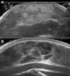
Infantile hemangioma. a Sonogram shows hyperechoic pattern, 8-month-old girl; b sonogram shows hypoechoic lobulated pattern, 5-month-old girl
Fig. 3
Fig. 3
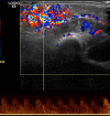
Color Doppler sonogram shows high vascular density, characteristic of infantile hemangioma
Fig. 4
Fig. 4
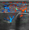
Infantile hemangioma in 4-month-old girl. B-flow technology shows the very high vascular density
Fig. 5
Fig. 5
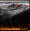
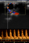
Infantile hemangioma. Spectral analysis shows arterial flow with low resistance and high velocities
Fig. 6
Fig. 6
Rapidly involuting congenital hemangioma (RICH) in 2-day-old boy (same case as of Fig. 8). Newborn with congenital vascular tumor in the occipital region. Biopsy confirmed the diagnosis
Fig. 7
Fig. 7
Rapidly involuting congenital hemangioma (RICH) in 1-month-old boy. Sonogram shows heterogeneous subcutaneous mass that contains large visible vessel (arrow)
Fig. 8
Fig. 8
Rapidly involuting congenital hemangioma (RICH) in 2-day-old boy. Sonogram shows heterogeneous subcutaneous mass with intralesional calcification (arrow) and large visible vessel (arrowhead)
Fig. 9
Fig. 9
Rapidly involuting congenital hemangioma (RICH) in 1-month-old boy. Color Doppler shows high vascular density, and spectral analysis shows the presence of low resistance arterial flow
Fig. 10
Fig. 10
Kaposiform hemangioendothelioma in a 2-day-old girl. The “extended view” sonogram shows a large subcutaneous mass of the abdominal wall (arrows). The mass is heterogeneous with some vessels visible on the gray-scale image. L liver, S spleen
Fig. 11
Fig. 11
Kaposiform hemangioendothelioma in a 2-day-old girl. Color Doppler sonogram confirms the high vascular density of lesion
Fig. 12
Fig. 12
Venous malformation. Compressible mass in the subcutaneous soft tissue (arrow). Note the dysplastic ectatic superficial veins (arrowheads) and the bluish discoloration of the overlying skin
Fig. 13
Fig. 13
Venous malformation in 13-year-old girl. Sonogram shows well-margined masses with a “spongiform” echostructure, which is hypoechoic in comparison to the surrounding tissues. The presence of intralesional phlebolith (arrow)
Fig. 14
Fig. 14
Venous malformation in 5-year-old boy. Spectral analysis shows a low-velocity flow with non-modulated spectrum
Fig. 15
Fig. 15
Macrocystic lymphatic malformation in 1-month-old boy. Sonogram shows multicystic mass in the thoracic subcutaneous tissue. The cystic spaces are anechoic and separated by septa
Fig. 16
Fig. 16
Macrocystic lymphatic malformation in 13-month-old boy. Sonogram shows multicystic mass in the neck subcutaneous tissue. The cystic spaces are hyperechoic from hemorrhage. A fluid-debris level is seen within a cyst (arrow)
Fig. 17
Fig. 17
Macrocystic lymphatic malformation in 1-month-old boy. Doppler sonogram shows flow that is confined to the septa. Spectral analysis shows arterial flow with high resistance
Fig. 18
Fig. 18
Arteriovenous malformation in 3-month-old boy. Hypoechoic subcutaneous lesion (*) with thin hyperechoic rim (arrows)
Fig. 19
Fig. 19
Arteriovenous malformation in 4-year-old boy. Color Doppler shows arterial flow with low resistance and high velocities
Fig. 20
Fig. 20
Spectral venous analysis. a Non-pulsatile venous flow in infantile hemangioma; b pulsatile venous flow in the arteriovenous malformation

Similar articles

Cited by

References

    1. Mulliken JB, Burrows PE, Fishman SJ. Mulliken and Young’s vascular anomalies—hemangiomas and malformation. Oxford Med Online. 2013
    1. Alibert JL. Nosologie naturelle ou les maladies du corp humain distribuees par familless. Paris: Caille and Ravier; 1817. pp. 349–351.
    1. Virchow R. Die krankhaften Geswulsthe. Berlin: Hirschwald; 1863. Angiome; pp. 306–425.
    1. Malan E. Vascular malformation (Angiodysplasias) Milan: Carlo Erba; 1974.
    1. Mulliken JB, Glowacki J. Hemangiomas and vascular malformations in infants and children: a classification based on endothelial characteristics. Plast Reconstr Surg. 1982;69:412–422. doi: 10.1097/00006534-198203000-00002. - DOI - PubMed