

Sox7 negatively regulates prostate-specific membrane antigen (PSMA) expression through PSMA-enhancer
- PMID: 30488457
- PMCID: PMC6344945
- DOI: 10.1002/pros.23743
Sox7 negatively regulates prostate-specific membrane antigen (PSMA) expression through PSMA-enhancer
Abstract
Background: PSMA expression in the prostate epithelium is controlled by a cis-element, PSMA enhancer (PSME). PSME contains multiple binding sites for Sox proteins, and in this study, we identified Sox7 protein as a negative regulator of PSMA expression through its interaction with PSME.
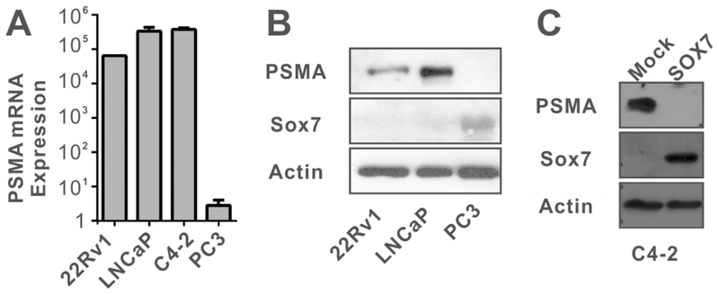
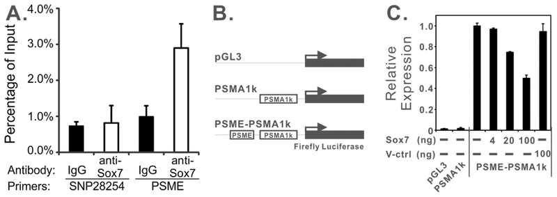
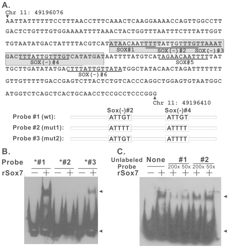
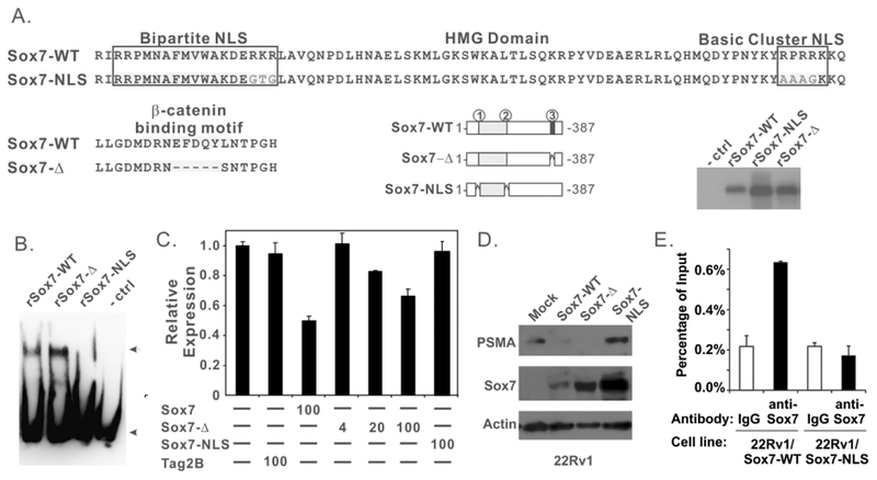
Methods: The statistical correlation between Sox7 and PSMA mRNA expression was evaluated using five prostate cancer studies from cBioportal. In vitro and in vivo interaction between Sox7 and PSME was evaluated by chromatin immunoprecipitation (ChIP), electrophoretic mobility shift assay (EMSA), and luciferase reporter assay. Synthetic oligonucleotides were generated to define the sites in PSME that interact with Sox7 protein. Sox7 mutants were generated to identify the region of this protein required to regulate PSMA expression. Sox7 was also stably expressed in LNCaP/C4-2 and 22Rv1 cells to validate the regulation of PSMA expression by Sox7 in vivo.
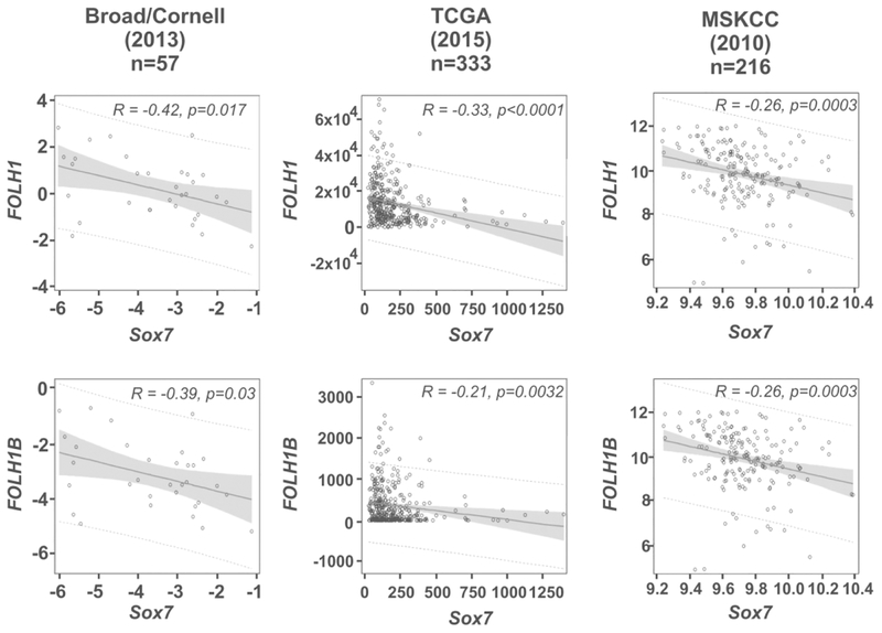
Results: Sox7 mRNA expression negatively correlated with PSMA/FOLH1 and PSMAL/FOLH1B mRNA expression in Broad/Cornell, TCGA and MSKCC studies, but not in two studies containing only metastatic prostate tumors. PC-3 cells mostly expressed the 48.5 KDa isoform 2 of Sox7, and the depletion of this isoform did not restore PSMA expression. Ectopic expression of canonical, wild-type Sox7 in C4-2 and 22Rv1 cells suppressed PSMA protein expression. ChIP assay revealed that canonical Sox7 protein preferentially interacts with PSME in vivo, and EMSA identified the SOX box sites #2 and #4 in PSME as required for its interaction. Sox7 was capable of directly binding to PSME and suppressed PSME-mediated transcription. The NLS regions of Sox7, but not its β-catenin interacting motif, are essential for this suppressing activity. Furthermore, restoration of wild-type Sox7 expression but not Sox7-NLS mutant in Sox7-null prostate cancer cell lines suppressed PSMA expression.
Conclusions: The inactivation of canonical Sox7 is responsible for the upregulated expression of PSMA in non-metastatic prostate cancer.
Keywords: WNT signaling; diagnostic and therapeutic marker; prostate-specific expression; transcriptional factor.
© 2018 Wiley Periodicals, Inc.
Conflict of interest statement
Disclosure Statement
Competing interests: The author(s) declare that they have no competing interests.
Figures
Similar articles
-
NFATc1 with AP-3 site binding specificity mediates gene expression of prostate-specific-membrane-antigen.J Mol Biol. 2003 Jul 18;330(4):749-60. doi: 10.1016/s0022-2836(03)00640-5. J Mol Biol. 2003. PMID: 12850144
-
Targeting prostate cancer with conditionally replicative adenovirus using PSMA enhancer.Mol Ther. 2004 Dec;10(6):1051-8. doi: 10.1016/j.ymthe.2004.08.028. Mol Ther. 2004. PMID: 15564137
-
A tissue-specific enhancer of the prostate-specific membrane antigen gene, FOLH1.Genomics. 2001 May 1;73(3):243-54. doi: 10.1006/geno.2000.6446. Genomics. 2001. PMID: 11350116
-
Tumor target prostate specific membrane antigen (PSMA) and its regulation in prostate cancer.J Cell Biochem. 2004 Feb 15;91(3):528-39. doi: 10.1002/jcb.10661. J Cell Biochem. 2004. PMID: 14755683 Review.
-
[Updated application of prostate-specific membrane antigen to the diagnosis and treatment of prostate cancer].Zhonghua Nan Ke Xue. 2008 Jan;14(1):79-82. Zhonghua Nan Ke Xue. 2008. PMID: 18297820 Review. Chinese.
Cited by
-
Reversible epigenetic alterations mediate PSMA expression heterogeneity in advanced metastatic prostate cancer.JCI Insight. 2023 Apr 10;8(7):e162907. doi: 10.1172/jci.insight.162907. JCI Insight. 2023. PMID: 36821396 Free PMC article.
-
Advances in PSMA theranostics.Transl Oncol. 2022 Aug;22:101450. doi: 10.1016/j.tranon.2022.101450. Epub 2022 May 18. Transl Oncol. 2022. PMID: 35597190 Free PMC article.
-
Membrane dual-targeting probes: A promising strategy for fluorescence-guided prostate cancer surgery and lymph node metastases detection.Acta Pharm Sin B. 2023 Mar;13(3):1204-1215. doi: 10.1016/j.apsb.2022.07.018. Epub 2022 Aug 3. Acta Pharm Sin B. 2023. PMID: 36970202 Free PMC article.
-
The clinical application of 68Ga-PSMA PET/CT and regulating mechanism of PSMA expression in patients with brain metastases of lung cancer.Transl Oncol. 2023 Feb;28:101616. doi: 10.1016/j.tranon.2023.101616. Epub 2023 Jan 6. Transl Oncol. 2023. PMID: 36621073 Free PMC article.
-
Prostate-Specific Membrane Antigen Expression and Response to DNA Damaging Agents in Prostate Cancer.Clin Cancer Res. 2022 Jul 15;28(14):3104-3115. doi: 10.1158/1078-0432.CCR-21-4531. Clin Cancer Res. 2022. PMID: 35552383 Free PMC article.
References
-
- Murphy GP, Barren RJ, Erickson SJ, et al. Evaluation and comparison of two new prostate carcinoma markers. Free-prostate specific antigen and prostate specific membrane antigen. Cancer. 1996;78(4):809–818. - PubMed
-
- Chang SS, Reuter VE, Heston WD, Bander NH, Grauer LS, Gaudin PB. Five different anti-prostate-specific membrane antigen (PSMA) antibodies confirm PSMA expression in tumor-associated neovasculature. Cancer Res. 1999;59(13):3192–3198. - PubMed
Publication types
MeSH terms
Substances
Grants and funding
LinkOut - more resources
Full Text Sources
Medical
Miscellaneous
