Two compartment pharmacokinetic model describes the intra-articular delivery and retention of rhprg4 following ACL transection in the Yucatan mini pig
- PMID: 30488470
- PMCID: PMC7201402
- DOI: 10.1002/jor.24191
Two compartment pharmacokinetic model describes the intra-articular delivery and retention of rhprg4 following ACL transection in the Yucatan mini pig
Abstract
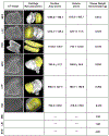
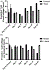
Treatment of the injured joint with rhPRG4 is based on recent observations that inflammation diminishes expression of native PRG4. Re-establishing lubrication between pressurized and sliding cartilage surfaces during locomotion promotes the nascent expression of PRG4 and thus intra-articular (IA) treatment strategies should be supported by pharmacokinetic evidence establishing the residence time of rhPRG4. A total of 21 Yucatan minipigs weighing ∼55 kg each received 4 mg of 131 I-rhPRG4 delivered by IA injection 5 days following surgical ACL transection. Animals were sequentially euthanized following IA rhPRG4 at 10 min (time zero), 24, 72 h, 6, 13 and 20 days later. The decay of the 131 I-rhPRG4 was measured relative to a non-injected aliquot and normalized to the weight of cartilage samples, menisci and synovium, and known cartilage volumes from each compartment surface obtained from representative Yucatan minipig knees. Decay of 131 I-rhPRG4 from joint tissues best fit a two-compartment model with an α half-life (t1/2α ) of 11.28 h and β half-life (t1/2β ) of 4.81 days. The tibial and femoral cartilage, meniscii, and synovium retained 7.7% of dose at 24 h. High concentrations of rhPRG4 were found in synovial fluid (SF) that was non-aspiratable and resided on the articular surfaces, removable by irrigation, at 10 min following 131 I-rhPRG4 injection. Synovial fluid K21 exceeded K12 and SF t1/2β was 28 days indicating SF is the reservoir for rhPRG4 following IA injection. Clinical Significance: rhPRG4 following IA delivery in a traumatized joint populates articular surfaces for a considerable period and may promote the native expression of PRG4. © 2018 Orthopaedic Research Society. Published by Wiley Periodicals, Inc. J Orthop Res 37:386-396, 2019.
Keywords: PRG4; cartilage; half-life; lubricin; pharmacokinetics.
© 2018 Orthopaedic Research Society. Published by Wiley Periodicals, Inc.
Conflict of interest statement
CONFLICTS OF INTEREST
G.D.J. and T.A.S. own equity in Lmbris BioPharma and have licensed patents related to the use of rhPRG4. T.S. also consults for Lμbris BioPharma.
Figures


Similar articles
-
Intra-articular Recombinant Human Proteoglycan 4 Mitigates Cartilage Damage After Destabilization of the Medial Meniscus in the Yucatan Minipig.Am J Sports Med. 2017 Jun;45(7):1512-1521. doi: 10.1177/0363546516686965. Epub 2017 Jan 27. Am J Sports Med. 2017. PMID: 28129516 Free PMC article.
-
Prevention of cartilage degeneration and restoration of chondroprotection by lubricin tribosupplementation in the rat following anterior cruciate ligament transection.Arthritis Rheum. 2010 Aug;62(8):2382-91. doi: 10.1002/art.27550. Arthritis Rheum. 2010. PMID: 20506144 Free PMC article.
-
Reduction of friction by recombinant human proteoglycan 4 in IL-1α stimulated bovine cartilage explants.J Orthop Res. 2017 Mar;35(3):580-589. doi: 10.1002/jor.23367. Epub 2017 Mar 2. J Orthop Res. 2017. PMID: 27411036 Free PMC article.
-
Quadruped Gait and Regulation of Apoptotic Factors in Tibiofemoral Joints following Intra-Articular rhPRG4 Injection in Prg4 Null Mice.Int J Mol Sci. 2022 Apr 12;23(8):4245. doi: 10.3390/ijms23084245. Int J Mol Sci. 2022. PMID: 35457064 Free PMC article.
-
Absence of Proteoglycan 4 (Prg4) Leads to Increased Subchondral Bone Porosity Which Can Be Mitigated Through Intra-Articular Injection of PRG4.J Orthop Res. 2019 Oct;37(10):2077-2088. doi: 10.1002/jor.24378. Epub 2019 Jun 26. J Orthop Res. 2019. PMID: 31119776
Cited by
-
Proteoglycan 4 (PRG4) treatment enhances wound closure and tissue regeneration.NPJ Regen Med. 2022 Jun 24;7(1):32. doi: 10.1038/s41536-022-00228-5. NPJ Regen Med. 2022. PMID: 35750773 Free PMC article.
-
Immunomodulatory Microparticles Epigenetically Modulate T Cells and Systemically Ameliorate Autoimmune Arthritis.Adv Sci (Weinh). 2023 Apr;10(11):e2202720. doi: 10.1002/advs.202202720. Epub 2023 Mar 8. Adv Sci (Weinh). 2023. PMID: 36890657 Free PMC article.
-
Recombinant manufacturing of multispecies biolubricants.bioRxiv [Preprint]. 2024 May 5:2024.05.05.592580. doi: 10.1101/2024.05.05.592580. bioRxiv. 2024. PMID: 38746339 Free PMC article. Preprint.
-
Delivery of FGF18 using mRNA-LNP protects the cartilage against degeneration via alleviating chondrocyte senescence.J Nanobiotechnology. 2025 Jan 22;23(1):34. doi: 10.1186/s12951-025-03103-9. J Nanobiotechnology. 2025. PMID: 39844298 Free PMC article.
-
Tryptase β regulation of joint lubrication and inflammation via proteoglycan-4 in osteoarthritis.Nat Commun. 2023 Apr 6;14(1):1910. doi: 10.1038/s41467-023-37598-3. Nat Commun. 2023. PMID: 37024468 Free PMC article.
References
Publication types
MeSH terms
Substances
Grants and funding
LinkOut - more resources
Full Text Sources
Miscellaneous

