. 2019 Jul;234(7):11587-11601.
doi: 10.1002/jcp.27791. Epub 2018 Nov 29.

Ranolazine prevents pressure overload-induced cardiac hypertrophy and heart failure by restoring aberrant Na+ and Ca2+ handling

Affiliations

Ranolazine prevents pressure overload-induced cardiac hypertrophy and heart failure by restoring aberrant Na+ and Ca2+ handling

Jiali Nie et al. J Cell Physiol. 2019 Jul.

Abstract

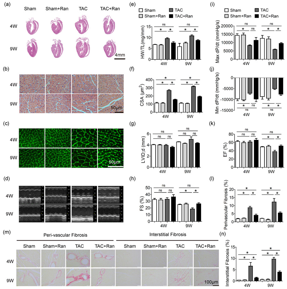
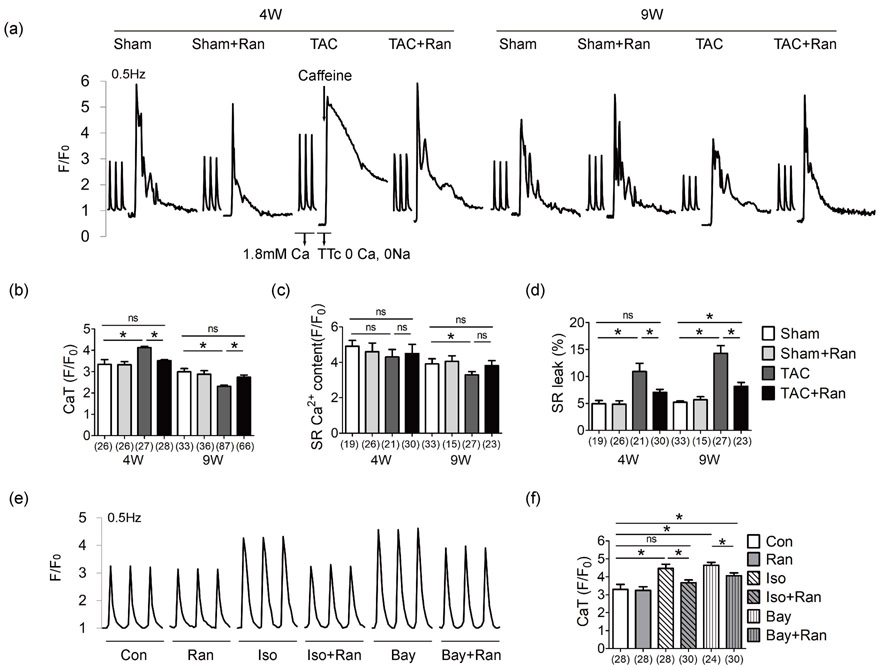
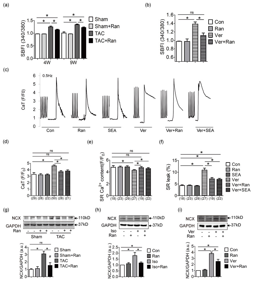
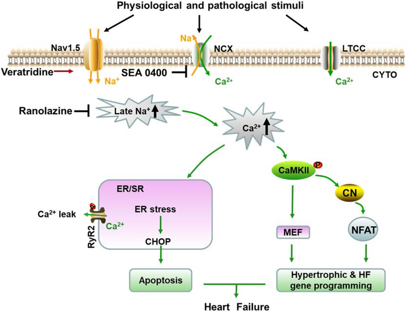
Background: Cardiac hypertrophy and heart failure are characterized by increased late sodium current and abnormal Ca2+ handling. Ranolazine, a selective inhibitor of the late sodium current, can reduce sodium accumulation and Ca 2+ overload. In this study, we investigated the effects of ranolazine on pressure overload-induced cardiac hypertrophy and heart failure in mice.

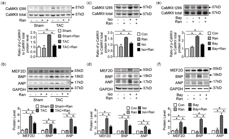
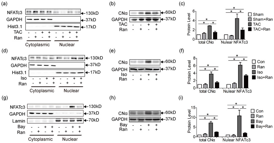
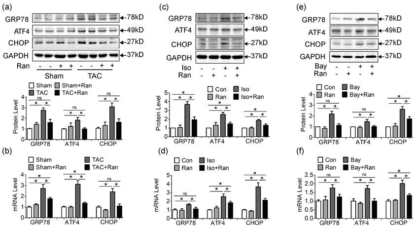
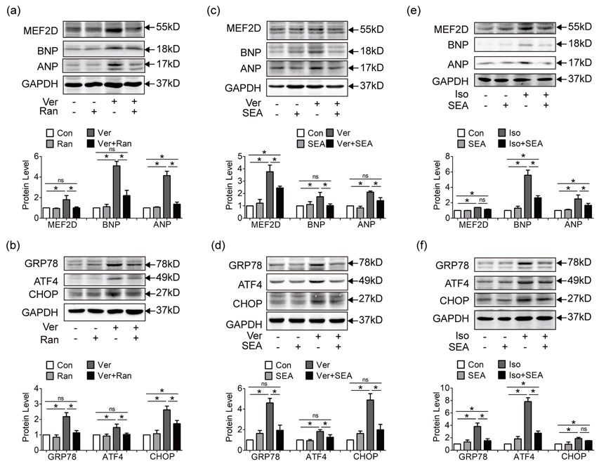
Methods and results: Inhibition of late sodium current with the selective inhibitor ranolazine suppressed cardiac hypertrophy and fibrosis and improved heart function assessed by echocardiography, hemodynamics, and histological analysis in mice exposed to chronic pressure overload induced by transverse aortic constriction (TAC). Ca2+ imaging of ventricular myocytes from TAC mice revealed both abnormal SR Ca 2+ release and increased SR Ca 2+ leak. Ranolazine restored aberrant SR Ca 2+ handling induced by pressure overload. Ranolazine also suppressed Na + overload induced in the failing heart, and restored Na + -induced Ca 2+ overload in an sodium-calcium exchanger (NCX)-dependent manner. Ranolazine suppressed the Ca 2+ -dependent calmodulin (CaM)/CaMKII/myocyte enhancer factor-2 (MEF2) and CaM/CaMKII/calcineurin/nuclear factor of activated T-cells (NFAT) hypertrophy signaling pathways triggered by pressure overload. Pressure overload also prolonged endoplasmic reticulum (ER) stress leading to ER-initiated apoptosis, while inhibition of late sodium current or NCX relieved ER stress and ER-initiated cardiomyocyte apoptosis.

Conclusions: Our study demonstrates that inhibition of late sodium current with ranolazine improves pressure overload-induced cardiac hypertrophy and systolic and diastolic function by restoring Na+ and Ca 2+ handling, inhibiting the downstream hypertrophic pathways and ER stress. Inhibition of late sodium current may provide a new treatment strategy for cardiac hypertrophy and heart failure.

Keywords: Ca2+ transient; Heart failure; INa,L; hypertrophy; ranolazine.


Figures

Figure 1
Figure 1
Ranolazine blunts pressure overload-induced cardiac hypertrophy and heart failure. (a) Examples of HE staining of heart sections. Examples of HE staining (b) and Wheat germ agglutinin staining (c) of heart cross-sections. (d) M-mode echocardiogram. (e) Statistics of HW/TL. (f) Statistics of CSA. (g) Statistics of LVID;d by echocardiography. (h) Statistics of FS by echocardiography. Quantification of max dP/dt, peak rate of pressure increase (i), and min dP/dt, peak rate of pressure decline (j), measured by aortic catheterization. (k) Statistics of EF by echocardiography. Sirius red stained myocardium section (m) and quantification (l and n) of cardiac fibrosis. Mice number was 5–14 per group. One-way ANOVA was performed for groups with the same intervention time; e.g., in (e), HW/TL values of Sham, Sham + Ran, TAC and TAC + Ran at 4 weeks were compared using one-way ANOVA, while HW/TL values of Sham, Sham + Ran, TAC and TAC + Ran at 9 weeks were compared using another one-way ANOVA independently. The same went for the rest of statistics. All data were shown as mean ± standard error of the mean (n ≥ 5; *p < 0.05). ANOVA: analysis of variance; CSA: cross sectional area; EF: ejection fraction; FS: fractional shortening; HW/TL: heart weight/tibia length; LVID;d: left ventricular internal diameter at diastole; Ran: ranolazine dyhydrocloride; TAC: transverse aortic constriction surgery; 4 W, 9 W: 4 weeks or 9 weeks after TAC operation
Figure 2
Figure 2
Ranolazine restores aberrant intracellular Ca2+ induced by pressure overload. (a) Representative Ca2+ recordings obtained in ventricular myocytes stimulated at 0.5 Hz. Ca2+ transient was recorded with Tyrode’s perfusion containing 1.8 mM Ca2+. SR leak was measured by switching the perfusion to 1 mM TTc buffer containing 0 Ca2+ and 0 Na+ to block SR leak, and caffeine was applied to empty SR Ca2+ storage. Quantification of CaT amplitude (b), SR Ca2+ content (c) and SR leak percentage (d). The Ca2+ transient recordings (e) and quantification (f) of the CaT amplitude in cardiomyocytes isolated from control C57/BL mice under different stimulus. Numbers indicate the number of cells for each condition (3–5 mice per group). Myocytes were concurrently treated with Bay/Iso and Ran 1 hr before the fluorescence detection. Ran, 10 μM; Iso, 20 μM; Bay, Bay K8644, LTCC activator, 1 μM. One-way ANOVA was performed for groups with the same intervention time; e.g., in (b), amplitude of CaT values of Sham, Sham + Ran, TAC and TAC + Ran at 4 weeks were compared using one-way ANOVA, while CaT values of Sham, Sham + Ran, TAC, and TAC + Ran at 9 weeks were compared using another one-way ANOVA independently. The same applied to (c,d). One-way ANOVA was performed for the analysis of (f). All data were shown as mean ± standard error of the mean (*p < 0.05). ANOVA: analysis of variance; Bay: Bay K8644; CaT: Ca2+ transient; Iso, isoproterenol hydrochloride; LTCC: L-type calcium channel; Ran: ranolazine dyhydrochloride; TAC: transverse aortic constriction surgery; TTc: tetracaine
Figure 3
Figure 3
Ranolazine suppresses Na+ induced Ca2+ overload via NCX in the failing heart. (a) Fluorescence ratio of SBFI-AM staining to mearure intracellular Na+ (3–5 mice per group). (b) Intracellular Na+ change in response to Ver and Ran treatment. Cardiomyocytes were isolated from control C57/BL mice (replicates n ≥ 5 per group). Myocytes were concurrently treated with Ver and Ran 1 hr before the SBFI-AM fluorescence detection. Representative Ca2+ recordings (c) and quantification of CaT amplitude (d), SR Ca2+ content (e) and SR leak percentage (f) of contol C57/BL mouse cardiomyocytes in response to Ver, Ran, and SEA. Drugs were given concurrently to myocytes for 1 hr before Fluo 4-AM fluorescence detection. Numbers indicate the number of cells for each condition. (g) NCX expression in mice after 4 weeks. NCX expression in Iso treated HL-1 cells (h) and Ver treated HL-1 cells (i). HL-1 cells were treated with Iso/Ver and Ran for 24 hr before protein extraction. Each western blot analysis was performed for 3–5 biological repeats. For (a) the 340/380 ratio of Sham, Sham + Ran, TAC and TAC + Ran at 4 weeks were compared using one-way ANOVA, while 340/380 ratio of Sham, Sham + Ran, TAC and TAC + Ran at 9 weeks were compared using another one-way ANOVA independently. One-way ANOVA was applied for the statistic of (b,d-i). All data were shown as mean ± standard error of the mean (*p < 0.05). ANOVA: analysis of variance; CaT: Ca2+ transient; Iso: isoproterenol hydrochloride; NCX: sodium calcium exchanger; Ran: ranolazine dyhydrochloride; SBFI-AM: sodium-binding benzofuran isophthalate-AM; SEA: SEA0400; TAC, transverse aortic constriction surgery; Ver: veratridine
Figure 4
Figure 4
Ranolazine inhibits Ca2+-dependent Ca2+/CaM/CaMKII/MEF2D hypertrophic pathway. (a, b) Western blot analysis of myocardium lysate at 4 weeks. CaMKII was highly phosphorylated in TAC mice, and the downstream hypertrophic markers MEF2D, ANP, and BNP were upregulated, while in ranolazine treated TAC mice, this pathway was inhibited. Hypertrophic markers were also tested in mouse cardiomyocyte cell line HL-1 exposed to Iso (c, d) or Bay (e, f). Each western blot analysis was performed for 3–5 biological repeats. One-way analysis of variance was used for analysis. Data were shown as mean ± standard error of the mean (*p < 0.05). ANP: atrial natriuretic peptide; Bay: Bay K8644; BNP: brain natriuretic peptide; CaMKII: Calcium/calmodulin-dependent protein kinase II; Iso: isoproterenol hydrochloride; MEF2D: myocyte enhancer factor 2D; TAC: transverse aortic constriction surgery
Figure 5
Figure 5
Ranolazine inhibits Ca2+ dependent Ca2+/calcineurin/NFAT hypertrophic pathway. (a) Cytoplasmic and nuclear proteins were extracted from mouse myocardium at 4 weeks for western blot analysis. GAPDH was taken as the cytoplasmic loading control. Histone H3 (Hist3.1) and Lamin B (Lamin) were taken as the nuclear loading control. Data showed that NFATc3 nuclear localization was increased in TAC mice and was significantly reduced by ranolazine. (b) Western blot analysis of whole cell lysate revealed increased expression of calcineurin catalytic α subunit (CNα), which was reduced by ranolazine. (c) showed statistics of (a) and (b). CNα was also upregulated in Iso (e) and Bay (h) treated HL-1 cells, followed by increased nuclear localization of NFATc3 (d,g), and treatment with ranolazine significantly inhibited the calcineurin/NFAT pathway. Statistics were shown in (f) and (i). Each western blot analysis was performed for 3–5 biological repeats. One-way analysis of variance was used for analysis. Data were shown as mean ± standard error of the mean (*p < 0.05). GAPDH: glyceraldehyde 3-phosphate dehydrogenase; NFAT: nuclear factor of activated T-cells; TAC: transverse aortic constriction surgery
Figure 6
Figure 6
Ranolazine suppresses ER stress induced by pressure overload and Ca2+ overload. (a, b) Both protein level (a) and mRNA level (b) of ER stress markers GRP78, ATF4 and CHOP were increased in TAC mice at 4 weeks, while ranolazine suppressed ER stress induced by pressure overload. (c, d) ER stress markers increased in Iso pretreated HL-1 cells, while ranolazine inhibited ER stress at both protein and mRNA levels. (e, f) ER stress markers increased in LTCC activator Bay K8644 pretreated HL-1 cells, and ranolazine also reduced such ER stress at both protein and mRNA levels. Each western blot analysis was performed for 3–5 biological repeats. Real-time PCR was performed for three biological replicates, and three technical repeats for each replicate. One-way analysis of variance was used for analysis. Data were shown as mean ± standard error of the mean (*p < 0.05). ATF4: activating transcription factor 4; CHOP: CCAAT-enhancer-binding protein homologous protein; ER: endoplasmic reticulum; GRP78: glucose-regulated protein 78; LTCC: L-type calcium channel; mRNA, messenger RNA; PCR: polymerase chain reaction
Figure 7
Figure 7
Inhibition of NCX reduces Ca2+-dependent hypertrophic pathway and ER stress. (a, b) Hypertrophic markers MEF2D, ANP, and BNP (a) and ER stress markers GRP78, ATF4, and CHOP (b) were upregulated in HL-1 pretreated with Na+ activator veratridine, while ranolazine suppressed hypertrophic pathway and ER stress induced by veratridine. (c, d) Hypertrophic markers (c) and ER stress markers (d) were upregulated in HL-1 pretreated with veratridine, while inhibition of NCX activity with SEA suppressed hypertrophic pathway and ER stress induced by veratridine. (e, f) Hypertrophic markers (e) and ER stress markers (f) were upregulated by β-agonist Isoproterenol in HL-1 cells, and inhibition of NCX activity with SEA also attenuated hypertrophy and ER stress. Each western blot analysis was performed for 3–5 biological repeats. Data were shown as mean ± standard error of the mean (*p < 0.05). ANP: atrial natriuretic peptide; ATF4: activating transcription factor 4; BNP: brain natriuretic peptide; CHOP: CCAAT-enhancer-binding protein homologous protein; ER: endoplasmic reticulum; GRP78: glucose-regulated protein 78; MEF2: myocyte enhancer factor 2; NCX: sodium calcium exchanger; SEA: SEA0400
Figure 8
Figure 8
Schematic showing how ranolazine attenuates cardiac hypertrophy and heart failure. Under physiological situation, INa,L constitutes a small inward sodium current, and NCX works in the forward mode to bring Na+ in and export Ca2+ out of the cell membrane. While under stimuli such as pressure overload, sustained INa,L makes up a considerable amount of Na+ current, and results in an elevated concentration of intracellular Na+, favoring the reverse mode of NCX to expel extra Na+ and bring in more Ca2+. Excessive Ca2+ triggers its target pathways and stress response. Under Ca2+ overload, Ca2+ leak from ER also increased, partly owing to the highly phosphorylated RyR2 by CaMKII, aggravating intracellular Ca2+ overload. In addition, imbalance of cytoplasmic and ER luminal Ca2+ handling disturb the protein folding environment and unfolded protein aggregates lead to ER stress. Prolonged ER stress up-regulates the apoptotic effector CHOP to induce cell apoptosis and heart failure. Activated CaMKII releases the transcriptional activity of transcription factor MEF2D and promote nuclear translocalization of NFATc3, allowing for hypertrophic and heart failure gene programming. Together, ranolazine exerts protection by inhibition of INa,L and normalizing Ca2+ handling, thus alleviating the downstream deterioration of cardiac structural and functional remodeling.

Similar articles

Cited by

References

    1. Armoundas AA, Hobai IA, Tomaselli GF, Winslow RL, and O’Rourke B. 2003. Role of sodium-calcium exchanger in modulating the action potential of ventricular myocytes from normal and failing hearts. Circulation research. 93:46–53. - PMC - PubMed
    1. Brill DM, and Wasserstrom JA. 1986. Intracellular sodium and the positive inotropic effect of veratridine and cardiac glycoside in sheep Purkinje fibers. Circulation research. 58:109–119. - PubMed
    1. Chaitman BR 2004. Efficacy and safety of a metabolic modulator drug in chronic stable angina: review of evidence from clinical trials. Journal of cardiovascular pharmacology and therapeutics. 9 Suppl 1:S47–64. - PubMed
    1. Chelu MG, Sarma S, Sood S, Wang S, van Oort RJ, Skapura DG, Li N, Santonastasi M, Muller FU, Schmitz W, Schotten U, Anderson ME, Valderrabano M, Dobrev D, and Wehrens XH. 2009. Calmodulin kinase II-mediated sarcoplasmic reticulum Ca2+ leak promotes atrial fibrillation in mice. The Journal of clinical investigation. 119:1940–1951. - PMC - PubMed
    1. Cingolani OH, Yang XP, Cavasin MA, and Carretero OA. 2003. Increased systolic performance with diastolic dysfunction in adult spontaneously hypertensive rats. Hypertension. 41:249–254. - PubMed

Publication types