. 2019 Feb;79(3):320-330.
doi: 10.1002/pros.23738. Epub 2018 Nov 28.

Tumor-infiltrating mesenchymal stem cells: Drivers of the immunosuppressive tumor microenvironment in prostate cancer?

Affiliations

Tumor-infiltrating mesenchymal stem cells: Drivers of the immunosuppressive tumor microenvironment in prostate cancer?

Timothy E Krueger et al. Prostate. 2019 Feb.

Abstract

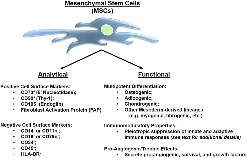
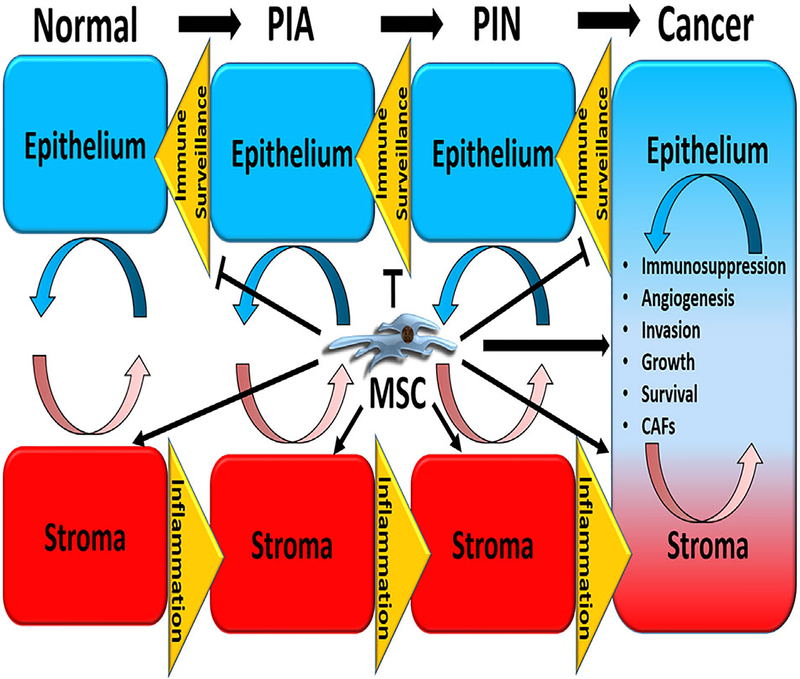
Background: Prostate cancer is characterized by T-cell exclusion, which is consistent with their poor responses to immunotherapy. In addition, T-cells restricted to the adjacent stroma and benign areas are characterized by anergic and immunosuppressive phenotypes. In order for immunotherapies to produce robust anti-tumor responses in prostate cancer, this exclusion barrier and immunosuppressive microenvironment must first be overcome. We have previously identified mesenchymal stem cells (MSCs) in primary and metastatic human prostate cancer tissue.

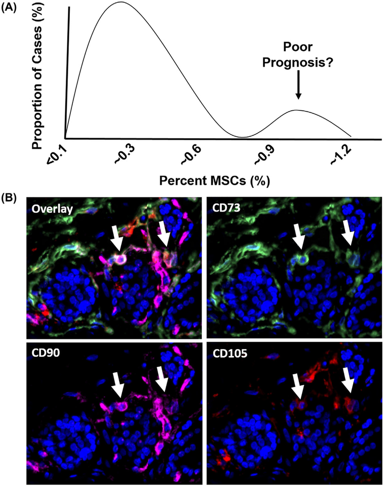
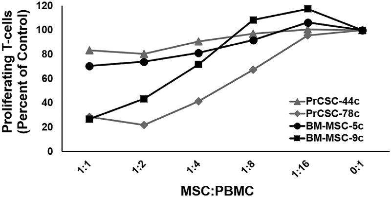
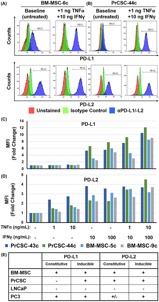
Methods: An Opal Multiplex immunofluorescence assay based on CD73, CD90, and CD105 staining was used to identify triple-labeled MSCs in human prostate cancer tissue. T-cell suppression assays and flow cytometry were used to demonstrate the immunosuppressive potential of primary MSCs expanded from human bone marrow and prostate cancer tissue from independent donors.

Results: Endogenous MSCs were confirmed to be present at sites of human prostate cancer. These prostate cancer-infiltrating MSCs suppress T-cell proliferation in a dose-dependent manner similar to their bone marrow-derived counterparts. Also similar to bone marrow-derived MSCs, prostate cancer-infiltrating MSCs upregulate expression of PD-L1 and PD-L2 on their cell surface in the presence of IFNγ and TNFα.

Conclusion: Prostate cancer-infiltrating MSCs suppress T-cell proliferation similar to canonical bone marrow-derived MSCs, which have well-documented immunosuppressive properties with numerous effects on both innate and adaptive immune system function. Thus, we hypothesize that selective depletion of MSCs infiltrating sites of prostate cancer should restore immunologic recognition and elimination of malignant cells via broad re-activation of cytotoxic pro-inflammatory pathways.

Keywords: MSC; T-cell exclusion; immunotherapy; mesenchymal stem cell; prostate cancer.


Conflict of interest statement

CONFLICT OF INTEREST

The authors have declared there are no relevant conflicts of interest to disclose.

Figures

FIGURE 1
FIGURE 1
Analytical and functional characterization of human mesenchymal stem cells. MSCs are defined by a series of positive and negative cell surface markers, in addition to functional properties, including multipotent differentiation potential, immunomodulatory properties, and pro-angiogenic/trophic effects
FIGURE 2
FIGURE 2
Potential roles of tumor-infiltrating mesenchymal stem cells in prostate cancer progression. Reciprocal interactions between the epithelium and stroma play important roles in diverse physiological and pathophysiological processes, including cancer initiation and progression. The latter is facilitated in part via inflammatory inducers and the suppression of immune surveillance. Tumor-infiltrating MSCs may play a role in cancer progression via multiple mechanisms, including stimulating angiogenesis, invasion, growth, survival, and the generation of carcinoma-associated fibroblasts (CAFs), in addition to suppression of innate and adaptive anti-tumor immune responses
FIGURE 3
FIGURE 3
Mesenchymal Stem Cells are present in human primary prostate cancer. A, Bimodal distribution pattern of MSCs in primary human prostate cancer based on multiparameter flow cytometry as previously described.39 B, Endogenous MSCs (white arrows) identified in human prostate cancer tissue based on positive staining for CD73 (green), CD90 (pink), and CD105 (red) using a multiplex immunofluorescence assay. Nuclei stained with DAPI (blue). 200× magnification
FIGURE 4
FIGURE 4
Human bone marrow-derived and tumor-infiltrating mesenchymal stem cells suppress T-cell proliferation. CellTrace Violet (CTV)-labeled PBMCs alone or with increasing ratios of “unlicensed” BM- or PCa-infiltrating MSCs (ie, PrCSCs) expanded from two independent donors each were used in a direct co-culture assay. Cultures incubated for 4 days in the presence of anti-CD3/-CD28 beads at 37°C, then collected for analysis by flow cytometry. T-cell proliferation defined as the number of CD3+ cells in the CTV-low population and calculated as a percentage of the stimulated PBMCs alone control (ie, 0:1)
FIGURE 5
FIGURE 5
Human bone marrow-derived and tumor-infiltrating mesenchymal stem cells upregulate PD-L1 and PD-L2 in response to IFNγ signaling. Representative flow plots documenting that MSCs expanded from human (A) bone marrow or (B) primary prostate cancer tissue (BM-MSC or PrCSC, respectively) upregulate PD-L1 and PD-L2 in response to IFNγ and TNFα. Median fluorescence intensity (MFI) of (C) PD-L1 and (D) PD-L2 expression on BM-MSCs and PrCSCs incubated in the presence or absence of IFNγ (10–100 ng/mL) and/or TNFα (1–10 ng/mL) for 24 h prior to staining for the respective marker or isotype control. PC3 and LNCaP used as controls to document baseline constitutive or inducible expression as previously reported.92 E, Summary of constitutive and inducible PD-L1 and PD-L2 expression

References

    1. Helmink BA, Gaudreau PO, Wargo JA. Immune Checkpoint Blockade across the Cancer Care Continuum. Immunity. 2018; 48:1077–1080. - PubMed
    1. June CH, Sadelain M. Chimeric Antigen Receptor Therapy. New Engl J Med. 2018; 379:64–73. - PMC - PubMed
    1. Schweizer MT, Drake CG. Immunotherapy for prostate cancer: recent developments and future challenges. Cancer Metastasis Rev. 2014; 33:641–655. - PMC - PubMed
    1. Slovin SF. Immunotherapy for prostate cancer: is prostate an immune responsive tumor? Curr Opin Urol. 2016;26:529–534. - PubMed
    1. Bilusic M, Madan RA, Gulley JL. Immunotherapy of Prostate Cancer: Facts and Hopes. Clin Cancer Res. 2017;23:6764–6770. - PMC - PubMed

Publication types