. 2019 Feb;110(2):639-649.
doi: 10.1111/cas.13893. Epub 2019 Jan 16.

Augmented antitumor activity of 5-fluorouracil by double knockdown of MDM4 and MDM2 in colon and gastric cancer cells

Affiliations

Augmented antitumor activity of 5-fluorouracil by double knockdown of MDM4 and MDM2 in colon and gastric cancer cells

Mamiko Imanishi et al. Cancer Sci. 2019 Feb.

Abstract

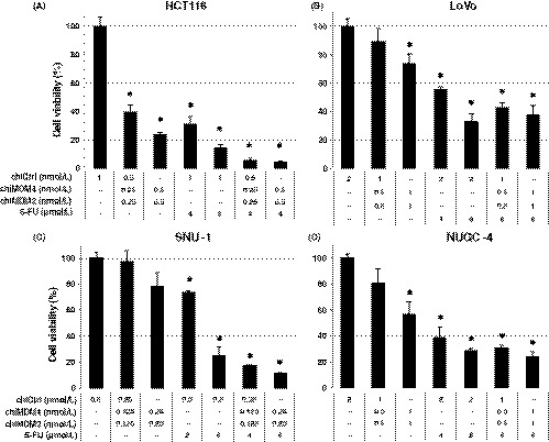
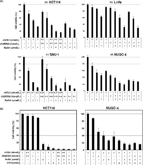
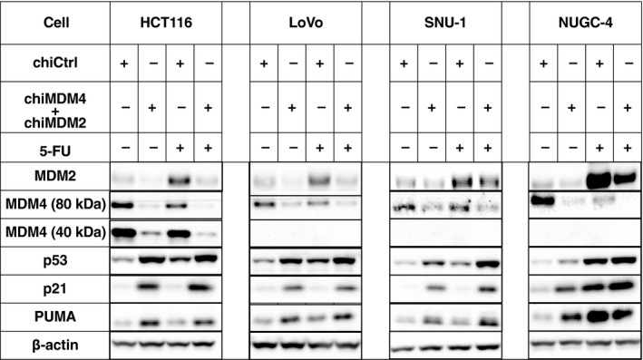
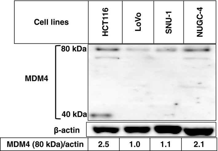
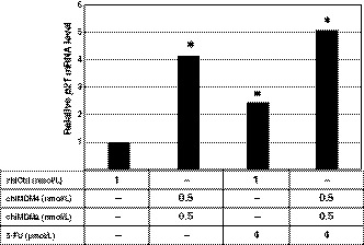
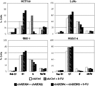
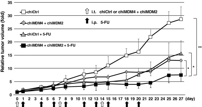
Inactivation of the TP53 tumor suppressor gene is essential during cancer development and progression. Mutations of TP53 are often missense and occur in various human cancers. In some fraction of wild-type (wt) TP53 tumors, p53 is inactivated by upregulated murine double minute homolog 2 (MDM2) and MDM4. We previously reported that simultaneous knockdown of MDM4 and MDM2 using synthetic DNA-modified siRNAs revived p53 activity and synergistically inhibited in vitro cell growth in cancer cells with wt TP53 and high MDM4 expression (wtTP53/highMDM4). In the present study, MDM4/MDM2 double knockdown with the siRNAs enhanced 5-fluorouracil (5-FU)-induced p53 activation, arrested the cell cycle at G1 phase, and potentiated the antitumor effect of 5-FU in wtTP53/highMDM4 human colon (HCT116 and LoVo) and gastric (SNU-1 and NUGC-4) cancer cells. Exposure to 5-FU alone induced MDM2 as well as p21 and PUMA by p53 activation. As p53-MDM2 forms a negative feedback loop, enhancement of the antitumor effect of 5-FU by MDM4/MDM2 double knockdown could be attributed to blocking of the feedback mechanism in addition to direct suppression of these p53 antagonists. Intratumor injection of the MDM4/MDM2 siRNAs suppressed in vivo tumor growth and boosted the antitumor effect of 5-FU in an athymic mouse xenograft model using HCT116 cells. These results suggest that a combination of MDM4/MDM2 knockdown and conventional cytotoxic drugs could be a promising treatment strategy for wtTP53/highMDM4 gastrointestinal cancers.

Keywords: 5-fluorouracil; MDM4; colon cancer; gastric cancer; p53.


Figures

Figure 1
Figure 1
Effects of double knockdown of MDM4 and MDM2 and 5‐fluorouracil (5‐FU) on the growth of colon (HCT116 and LoVo) and gastric cancer cell lines (SNU‐1 and NUGC‐4) with wtTP53/high MDM4. HCT116 (A), LoVo (B), SNU‐1 (C), and NUGC‐4 cells (D) were transfected with either DNA‐modified control siRNA (chiCtrl) or mixture of DNA‐modified siRNA targeting MDM4 (chiMDM4) and MDM2 (chiMDM2). After 4‐16 hours of incubation, cells were exposed to 5‐FU at the indicated concentrations. Five days after transfection, cell viability was determined using the WST‐8 assay. Cell viability relative to those transfected with chiCtrl are shown (mean ± SD; n = 3) . *P < .05, compared with the chiCtrl
Figure 2
Figure 2
Effects of MDM4 knockdown and nutlin‐3 on tumor cell growth and antitumor activity of 5‐fluorouracil (5‐FU) in colon and gastric cancer cells. A, Growth inhibitory effect of MDM4 knockdown and nutlin‐3 in HCT116 (a), LoVo (b), SNU‐1 (c), and NUGC‐4 cells (d). Cells were transfected with either control siRNA (chiCtrl) or DNA‐modified siRNA targeting MDM4 (chiMDM4). After 4‐16 hours of incubation, cells were exposed to nutlin‐3 at the indicated concentrations. Five days after transfection, cell viability was determined using the WST‐8 assay. Cell viability relative to those transfected with chiCtrl are shown (mean ± SD; n = 3). B, Enhancement of MDM4 knockdown/nutlin‐3 on antitumor activity of 5‐FU in colon (HCT116) and gastric cancer cells (NUGC‐4). HCT116 (left) and NUGC‐4 cells (right) were transfected with either chiCtrl or chiMDM4. After 16 hours of incubation, cells were exposed to nutlin‐3 and 5‐FU at the indicated concentrations. Five days after transfection, cell viability was determined using the WST‐8 assay. Cell viability relative to those transfected with chiCtrl are shown (mean ± SD; n = 3)
Figure 3
Figure 3
Effects of double knockdown of MDM2 and MDM4 and 5‐fluorouracil (5‐FU) on levels of p53, p21, and p53 upregulated modulator of apoptosis (PUMA) in colon (HCT116 and LoVo) and gastric cancer cells (SNU‐1 and NUGC‐4). HCT116, LoVo, SNU‐1, and NUGC‐4 cells were transfected with either control siRNA (chiCtrl) or a mixture of DNA‐modified siRNA targeting MDM4 (chiMDM4) and MDM2 (chiMDM2). After 4‐16 hours of incubation, cells were exposed to 5‐FU at the indicated concentrations. Twenty‐four hours after exposure to 5‐FU, cells were analyzed for levels of MDM2, MDM4, p53, p21, and PUMA using immunoblotting. β‐actin was used as an internal control
Figure 4
Figure 4
Expression level of MDM4 in wtTP53 colon (HCT116 and LoVo) and gastric cancer cell lines (SNU‐1 and NUGC‐4). Expression levels of MDM4 were analyzed by immunoblotting
Figure 5
Figure 5
Effects of double knockdown of MDM4 and MDM2 and 5‐fluorouracil (5‐FU) on the level of p21 mRNA in HCT116 cells. HCT116 cells were transfected with either control siRNA (chiCtrl) or a mixture of DNA‐modified siRNA targeting MDM4 (chiMDM4) and MDM2 (chiMDM2) for 16 hours and then cultured in the presence of 5‐FU. Forty‐eight hours after transfection, the cells were analyzed for their p21 mRNA level using quantitative RTPCR. p21 mRNA levels relative to those transfected with chiCtrl are shown. *P < .05, compared with the chiCtrl
Figure 6
Figure 6
Effects of double knockdown of MDM4 and MDM2 and 5‐fluorouracil (5‐FU) on cell cycle distribution of colon (HCT116 and LoVo) and gastric cancer cells (SNU‐1 and NUGC‐4). HCT116 (top left), LoVo (top right), SNU‐1 (bottom left), and NUGC‐4 cells (bottom right) were transfected with control siRNA (chiCtrl) or a mixture of DNA‐modified siRNA targeting MDM4 (chiMDM4) and MDM2 (chiMDM2) overnight, then exposed to 5‐FU. After overnight cultivation, the cells were analyzed for cell cycle distribution by flow cytometry
Figure 7
Figure 7
In vivo antitumor activity of a mixture of DNA‐modified siRNA targeting MDM4 (chiMDM4) and MDM2 (chiMDM2), 5‐fluorouracil (5‐FU), and combinations of these two in a xenograft model of HCT116 colon cancer cells. Results are expressed as means ± SE. *< .05, **< .001, compared with the control group (those injected with control siRNA [chiCtrl])

Similar articles

Cited by

References

    1. Douillard JY, Cunningham D, Roth AD, et al. Irinotecan combined with fluorouracil compared with fluorouracil alone as first‐line treatment for metastatic colorectal cancer: a multicentre randomised trial. Lancet. 2000;355:1041‐1047. - PubMed
    1. Saltz LB, Cox JV, Blanke C, et al. Irinotecan plus fluorouracil and leucovorin for metastatic colorectal cancer. Irinotecan Study Group. N Engl J Med. 2000;343:905‐914. - PubMed
    1. de Gramont A, Figer A, Seymour M, et al. Leucovorin and fluorouracil with or without oxaliplatin as first‐line treatment in advanced colorectal cancer. J Clin Oncol. 2000;18:2938‐2947. - PubMed
    1. Goldberg RM, Sargent DJ, Morton RF, et al. A randomized controlled trial of fluorouracil plus leucovorin, irinotecan, and oxaliplatin combinations in patients with previously untreated metastatic colorectal cancer. J Clin Oncol. 2004;22:23‐30. - PubMed
    1. Falcone A, Ricci S, Brunetti I, et al. Phase III trial of infusional fluorouracil, leucovorin, oxaliplatin, and irinotecan (FOLFOXIRI) compared with infusional fluorouracil, leucovorin, and irinotecan (FOLFIRI) as first‐line treatment for metastatic colorectal cancer: the Gruppo Oncologico Nord Ovest. J Clin Oncol. 2007;25:1670‐1676. - PubMed

MeSH terms