. 2019 Jan;9(1):e01168.
doi: 10.1002/brb3.1168. Epub 2018 Nov 28.

Resting-state functional brain networks in adults with a new diagnosis of focal epilepsy

Affiliations

Resting-state functional brain networks in adults with a new diagnosis of focal epilepsy

Batil K Alonazi et al. Brain Behav. 2019 Jan.

Abstract

Objectives: Newly diagnosed focal epilepsy (NDfE) is rarely studied, particularly using advanced neuroimaging techniques. Many patients with NDfE experience cognitive impairments, particularly with respect to memory, sustained attention, mental flexibility, and executive functioning. Cognitive impairments have been related to alterations in resting-state functional brain networks in patients with neurological disorders. In the present study, we investigated whether patients with NDfE had altered connectivity in large-scale functional networks using resting-state functional MRI.

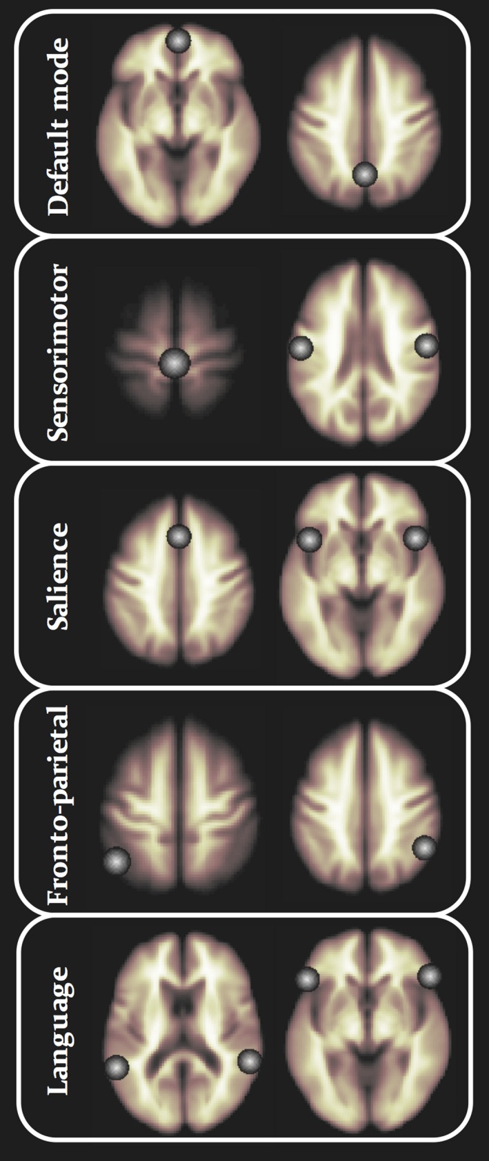
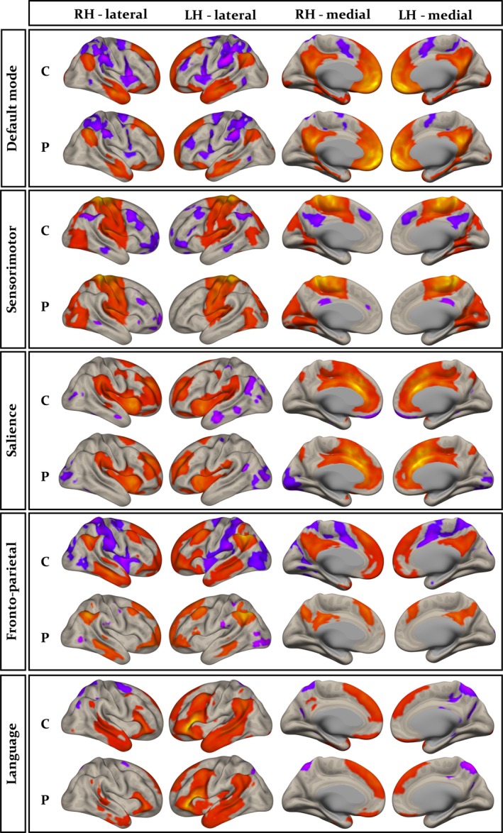
Methods: We recruited 27 adults with NDfE and 36 age- and sex-matched healthy controls. Resting-state functional MRI was analyzed using the Functional Connectivity Toolbox (CONN). We investigate reproducibly determined large-scale functional networks, including the default mode, salience, fronto-parietal attention, sensorimotor, and language networks using a seed-based approach. Network comparisons between patients and controls were thresholded using a FDR cluster-level correction approach.

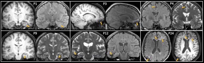
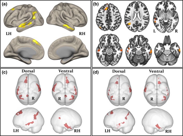
Results: We found no significant differences in functional connectivity between seeds within the default mode, salience, sensorimotor, and language networks and other regions of the brain between patients and controls. However, patients with NDfE had significantly reduced connectivity between intraparietal seeds within the fronto-parietal attention network and predominantly frontal and temporal cortical regions relative to controls; this finding was demonstrated including and excluding the patients with brain lesions. No common alteration in brain structure was observed in patients using voxel-based morphometry. Findings were not influenced by treatment outcome at 1 year.

Conclusions: Patients with focal epilepsy have brain functional connectivity alterations at diagnosis. Functional brain abnormalities are not necessarily a consequence of the chronicity of epilepsy and are present when seizures first emerge.

Keywords: brain connectivity; cognitive dysfunction; new-onset seizures; treatment outcome.


Figures

Figure 1
Figure 1
Location of seeds for each resting‐state network. See Table 2 for anatomical locations and coordinates
Figure 2
Figure 2
Lesions identified in the present study (see Table 1 for corresponding information). Patient 1 (P1): mesial temporal focal cortical dysplasia and atrophy of ipsilateral hippocampal head on T1‐weighted (left) and T2‐FLAIR (right) images; P3: orbitofrontal gliosis on T1‐weighted (left) and T2‐FLAIR (right) images; P4: focal cortical dysplasia of middle frontal gyrus on T2‐FLAIR (left) and T2‐weighted (right) images; P9: unilateral hippocampal atrophy on T1‐weighted (left) and T2‐weighted (right) images; P12: temporal lobe white matter alteration on T2‐weighted (left) and T2‐FLAIR (right) images; P24: frontal lobe gliosis and encephalomalacia, corpus callosum atrophy and contrecoup posterior gliosis on T2‐FLAIR (left) and T1‐weighted (right images). Patient 21 (slight unilateral hippocampal alteration) not illustrated. Images are neurological convention (right = right)
Figure 3
Figure 3
Resting‐state functional networks shown separately for controls (C) and patients (P). Regions correlated (orange) and anticorrelated (purple) with seeds are indicated. Specific seeds used to generate networks indicated here include medial prefrontal cortex (default mode), primary motor area (sensorimotor), anterior cingulate gyrus (salience), left intraparietal sulcus (fronto‐parietal), and left inferior frontal gyrus (language). Networks were reproducibly reconstructed using the alternative seeds shown in Figure 1. Note the visual difference between controls and patients in the fronto‐parietal attentional network
Figure 4
Figure 4
Significantly reduced functional connectivity within the fronto‐parietal attentional network in patients relative to controls (left intraparietal sulcus seed). Hypoconnectivity in all patients relative to controls is projected onto a 3D rendering (a) and axial sections (b) to illustrate anatomical locations. The spatial distribution of hypoconnectivity in all patients (c) and patients with normal MRI scans (d) is compared using glass brain projections. The corresponding information for each cluster is provided in Tables 3 and 4

Similar articles

Cited by

References

    1. Aikia, M. , Kalviainen, R. , Mervaala, E. , & Riekkinen, P. J., Sr. (1999). Predictors of seizure outcome in newly diagnosed partial epilepsy: Memory performance as a prognostic factor. Epilepsy Research, 37, 159–167. 10.1016/S0920-1211(99)00059-5 - DOI - PubMed
    1. Aikia, M. , Kalviainen, R. , & Riekkinen, P. J. (1995). Verbal learning and memory in newly diagnosed partial epilepsy. Epilepsy Research, 22, 157–164. 10.1016/0920-1211(95)00042-9 - DOI - PubMed
    1. Aikia, M. , Salmenpera, T. , Partanen, K. , & Kalviainen, R. (2001). Verbal memory in newly diagnosed patients and patients with chronic left temporal lobe epilepsy. Epilepsy and Behavior, 2, 20–27. 10.1006/ebeh.2000.0140 - DOI - PubMed
    1. Andrews‐Hanna, J. R. , Smallwood, J. , & Spreng, R. N. (2014). The default network and self‐generated thought: Component processes, dynamic control, and clinical relevance. Annals of the New York Academy of Sciences, 1316, 29–52. - PMC - PubMed
    1. Annegers, J. F. , Hauser, W. A. , & Elveback, L. R. (1979). Remission of seizures and relapse in patients with epilepsy. Epilepsia, 20, 729–737. 10.1111/j.1528-1157.1979.tb04857.x - DOI - PubMed

Publication types