

Upregulation of long non-coding RNA NNT-AS1 promotes osteosarcoma progression by inhibiting the tumor suppressive miR-320a
- PMID: 30489194
- PMCID: PMC6422455
- DOI: 10.1080/15384047.2018.1538612
Upregulation of long non-coding RNA NNT-AS1 promotes osteosarcoma progression by inhibiting the tumor suppressive miR-320a
Abstract
Objective: To investigate the role and mechanism of action of nicotinamide nucleotide transhydrogenase antisense RNA 1 (NNT-AS1) in osteosarcoma (OS).
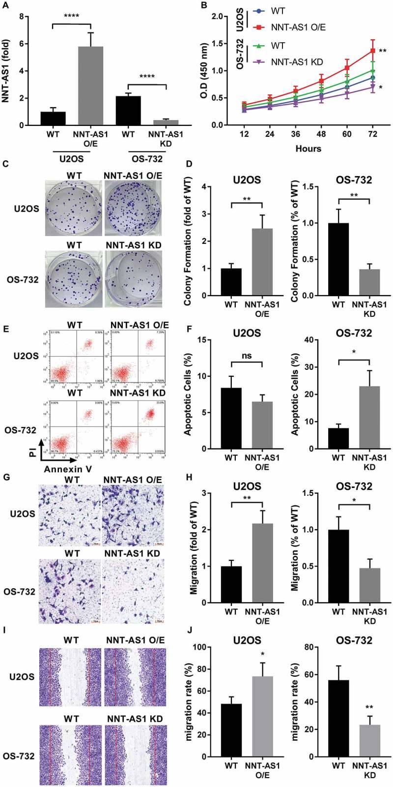
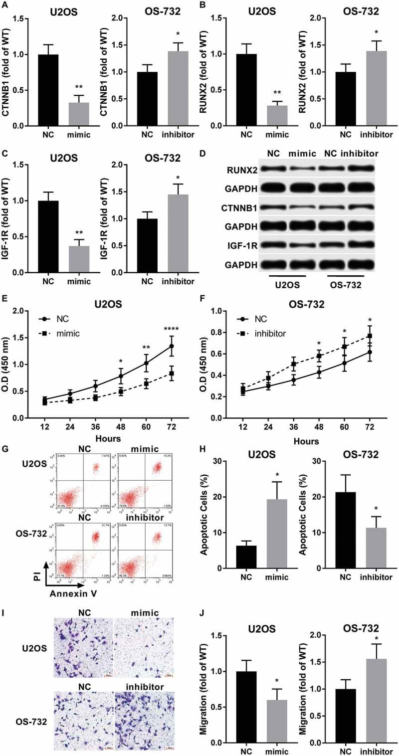
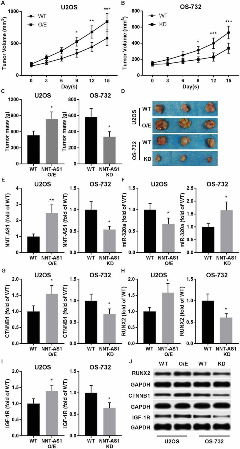
Methods: Bioinformatic analysis suggested miR-320a as potential target of NNT-AS1. Influence of NNT-AS1 overexpression or knockdown on OS cell proliferation, colony-formation, apoptosis, migration and invasion capacity was first investigated. Expression levels of NNT-AS1, miR-320a, beta-catenin, RUNX2, IGF-1R, c-Myc, Cyclin D1 and MMP13 were also evaluated by RT-qPCR and western blotting accordingly. Xenograft models using U2OS and OS-732 cells with different NNT-AS1 gene modifications were constructed for tumor formation assay as well as evaluation of miR-320a, beta-catenin and RUNX2 expression in primary lesion. NNT-AS1-overexpressing U2OS cells and NNT-AS1-knockdown OS-732 cells were subject to miR-320a mimic and inhibitor transfection, respectively, to investigate the miR-320a dependency of the osteosarcoma-promoting role of NNT-AS1.
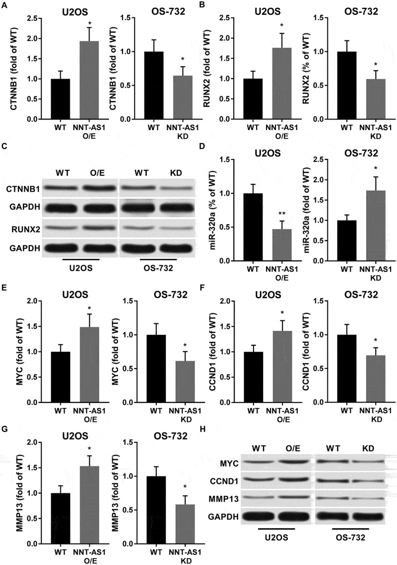
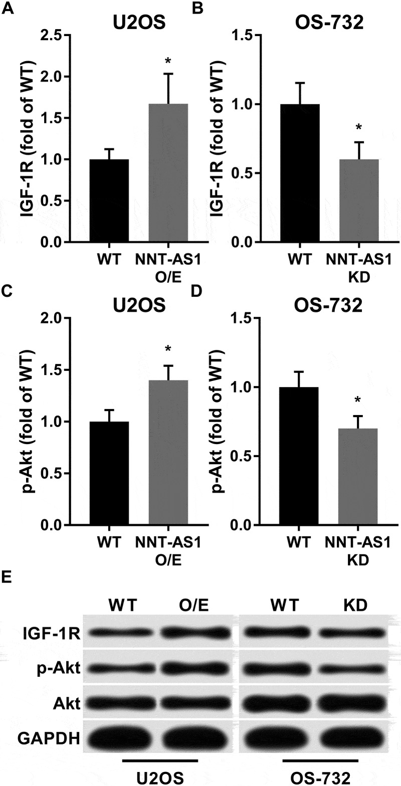
Results: NNT-AS1 overexpression significantly increased proliferation, survival and mobility of U2OS cells in vitro as well as its tumor formation ability in vivo, while NNT-AS1 knockdown showed opposite effect on OS-732 cells. In both in vitro and in vivo model, NNT-AS1 expression level significantly correlated with that of beta-catenin, RUNX2, IGF-1R, c-Myc, Cyclin D1 and MMP13 as well as Akt phosphorylation level, and inversely correlated with miR-320a expression. Transfection of miR-320a mimic significantly inhibiter the promoting effect of NNT-AS1 on cell proliferation, survival and mobility of U2OS cells, while miR-320 inhibitor partially rescued that of OS-732 cells.
Conclusion: NNT-As1 functions as a cancer-promoting lncRNA by downregulating miR-320a, thus increasing the protein expression level of beta-catenin, RUNX2 and IGF-1R as well as activation of Akt in osteosarcoma.
Keywords: NNT-AS1; long noncoding RNA; miR-320a; osteosarcoma.
Figures
Similar articles
-
LncRNA NNT-AS1 promotes non-small cell lung cancer progression through regulating miR-22-3p/YAP1 axis.Thorac Cancer. 2020 Mar;11(3):549-560. doi: 10.1111/1759-7714.13280. Epub 2020 Jan 10. Thorac Cancer. 2020. PMID: 31923353 Free PMC article.
-
Long noncoding RNA NNT-AS1 promotes gastric cancer proliferation and invasion by regulating microRNA-363 expression.J Cell Biochem. 2019 Apr;120(4):5704-5712. doi: 10.1002/jcb.27855. Epub 2018 Oct 15. J Cell Biochem. 2019. PMID: 30324628
-
Long non-coding RNA NNT-AS1 promotes cholangiocarcinoma cells proliferation and epithelial-to-mesenchymal transition through down-regulating miR-203.Aging (Albany NY). 2020 Feb 5;12(3):2333-2346. doi: 10.18632/aging.102747. Epub 2020 Feb 5. Aging (Albany NY). 2020. PMID: 32019904 Free PMC article.
-
Targeting the Wnt/β-catenin cascade in osteosarcoma: The potential of ncRNAs as biomarkers and therapeutics.Pathol Res Pract. 2024 Jul;259:155346. doi: 10.1016/j.prp.2024.155346. Epub 2024 May 11. Pathol Res Pract. 2024. PMID: 38781762 Review.
-
Insights on the potential oncogenic impact of long non-coding RNA nicotinamide nucleotide transhydrogenase antisense RNA 1 in different cancer types; integrating pathway(s) and clinical outcome(s) association.Pathol Res Pract. 2022 Dec;240:154183. doi: 10.1016/j.prp.2022.154183. Epub 2022 Oct 20. Pathol Res Pract. 2022. PMID: 36327824 Review.
Cited by
-
High expression of long non-coding RNA NNT-AS1 facilitates progression of cholangiocarcinoma through promoting epithelial-mesenchymal transition.Am J Transl Res. 2019 Sep 15;11(9):5438-5456. eCollection 2019. Am J Transl Res. 2019. PMID: 31632521 Free PMC article.
-
Hydroxygenkwanin suppresses proliferation, invasion and migration of osteosarcoma cells via the miR‑320a/SOX9 axis.Mol Med Rep. 2022 Oct;26(4):299. doi: 10.3892/mmr.2022.12815. Epub 2022 Aug 5. Mol Med Rep. 2022. PMID: 35929504 Free PMC article.
-
Serum long non‑coding RNA NNT‑AS1 protected by exosome is a potential biomarker and functions as an oncogene via the miR‑496/RAP2C axis in colorectal cancer.Mol Med Rep. 2021 Aug;24(2):585. doi: 10.3892/mmr.2021.12224. Epub 2021 Jun 16. Mol Med Rep. 2021. PMID: 34132374 Free PMC article.
-
Deciphering microRNA-mRNA regulatory network in adult T-cell leukemia/lymphoma; the battle between oncogenes and anti-oncogenes.PLoS One. 2021 Feb 25;16(2):e0247713. doi: 10.1371/journal.pone.0247713. eCollection 2021. PLoS One. 2021. PMID: 33630973 Free PMC article.
-
MicroRNA-320a enhances LRWD1 expression through the AGO2/FXR1-dependent pathway to affect cell behaviors and the oxidative stress response in human testicular embryonic carcinoma cells.Aging (Albany NY). 2024 Feb 21;16(4):3973-3988. doi: 10.18632/aging.205571. Epub 2024 Feb 21. Aging (Albany NY). 2024. PMID: 38385979 Free PMC article.
References
Publication types
MeSH terms
Substances
LinkOut - more resources
Full Text Sources
Medical
Research Materials
Miscellaneous