

Novel PLAG1 Gene Rearrangement Distinguishes a Subset of Uterine Myxoid Leiomyosarcoma From Other Uterine Myxoid Mesenchymal Tumors
- PMID: 30489320
- PMCID: PMC6764762
- DOI: 10.1097/PAS.0000000000001196
Novel PLAG1 Gene Rearrangement Distinguishes a Subset of Uterine Myxoid Leiomyosarcoma From Other Uterine Myxoid Mesenchymal Tumors
Abstract
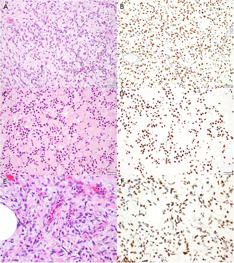
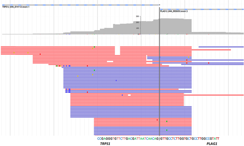
Genetic alterations in uterine myxoid leiomyosarcoma are unknown. We investigate the clinicopathologic features of 19 uterine tumors previously diagnosed as myxoid leiomyosarcomas in which tumoral RNA was subjected to targeted RNA sequencing. PLAG1, BCOR, BCORL1, HMGA2, and ALK break-apart fluorescence in situ hybridization (FISH) and BCOR, PLAG1, and ALK immunohistochemistry were performed in cases which failed or lacked fusions by sequencing. The diagnosis of myxoid leiomyosarcoma was confirmed in 15 cases after exclusion of 4 tumors with BCOR and ALK rearrangements. These 15 patients presented at a median age of 50 years with stage I (3), II (2), III (2), and IV (1) tumors, respectively; stage was unknown in 7 cases. Tumor size ranged from 10 to 24 cm. Matrix was myxoid in all tumors and also eosinophilic in 2. Cells were spindled, epithelioid, and both in 10, 2, and 3 tumors and showed mild, moderate, and severe nuclear atypia in 3, 8, and 4 tumors, respectively. Mitotic index ranged from <1 to 14/10 HPF, while tumor necrosis was present in 6 (40%). Novel TRPS1-PLAG1 or RAD51B-PLAG1 fusions were detected by sequencing in 4 tumors, 3 of which were also confirmed by FISH. Diffuse PLAG1 expression was seen in 7 tumors, including 4 with PLAG1 rearrangement. No morphologic differences were seen among PLAG1 fusion-positive and fusion-negative tumors. No PLAG1, HMGA2, ALK, BCOR, or BCORL1 rearrangements were detected by FISH in 11 tumors. On the basis of sequencing and FISH results, PLAG1 rearrangements resulting in PLAG1 expression underpin ~25% of myxoid leiomyosarcomas and may serve as a useful diagnostic biomarker. Immunohistochemistry, targeted RNA sequencing, and/or FISH may distinguish myxoid leiomyosarcoma from its morphologic mimics.
Conflict of interest statement
CONFLICTS OF INTEREST
The authors have no conflicts of interest to declare.
Figures
References
-
- King ME, Dickersin GR, Scully RE. Myxoid leiomyosarcoma of the uterus. A report of six cases. Am J Surg Pathol. 1982;6:589–98. - PubMed
-
- Abeler VM, Royne O, Thoresen S, et al. Uterine sarcomas in Norway. A histopathological and prognostic survey of a total population from 1970 to 2000 including 419 patients. Histopathology. 2009;54:355–64. - PubMed
-
- Burch DM, Tavassoli FA. Myxoid leiomyosarcoma of the uterus. Histopathology. 2011;59:1144–55. - PubMed
-
- Parra-Herran C, Schoolmeester JK, Yuan L, et al. Myxoid leiomyosarcoma of the uterus: a clinicopathologic analysis of 30 cases and review of the literature with reappraisal of its distinction from other uterine myxoid mesenchymal neoplasms. Am J Surg Pathol. 2016;40:285–301. - PubMed
-
- Bennett JA, Nardi V, Rouzbahman M, et al. Inflammatory myofibroblastic tumor of the uterus: a clinicopathological, immunohistochemical, and molecular analysis of 13 cases highlighting their broad morphologic spectrum. Mod Pathol. 2017;30:1489–503. - PubMed
MeSH terms
Substances
Grants and funding
LinkOut - more resources
Full Text Sources
Medical
