Clinical Trial
. 2019 Feb/Mar;42(2):65-72.
doi: 10.1097/CJI.0000000000000250.

Natural Killer Cell Homing and Persistence in the Bone Marrow After Adoptive Immunotherapy Correlates With Better Leukemia Control

Affiliations
Clinical Trial

Natural Killer Cell Homing and Persistence in the Bone Marrow After Adoptive Immunotherapy Correlates With Better Leukemia Control

Bartosz Grzywacz et al. J Immunother. 2019 Feb/Mar.

Abstract

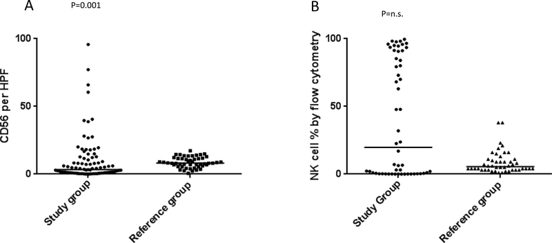
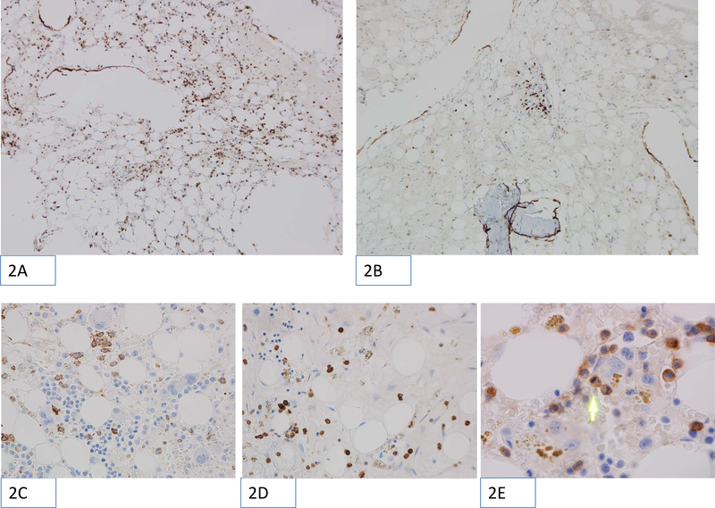
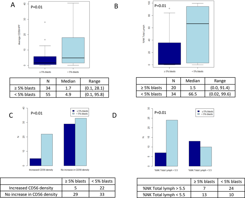
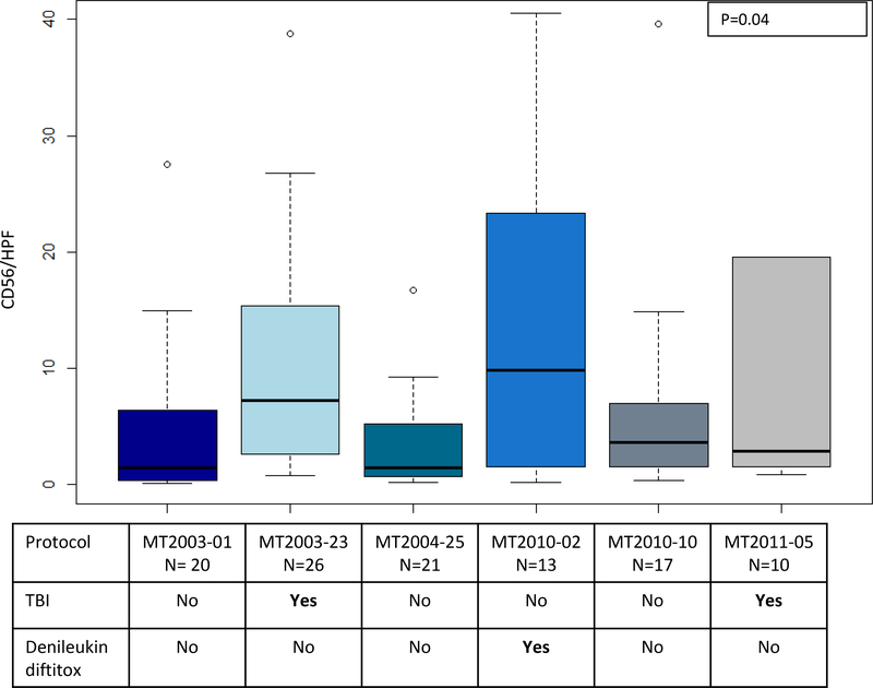
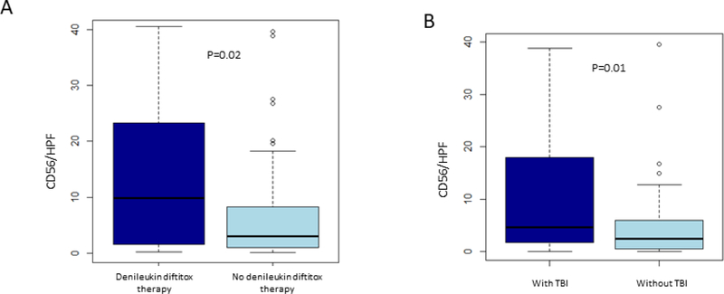
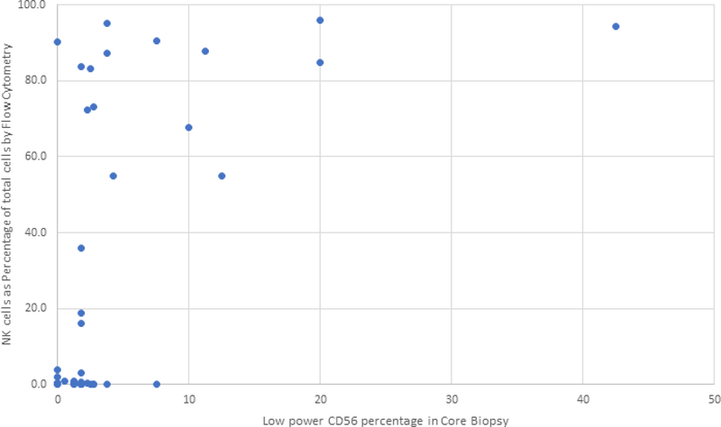
Cellular immunotherapy using allogeneic natural killer (NK) cells may overcome chemotherapy-refractory acute myeloid leukemia. Our goal was to document NK cell homing/persistence in the bone marrow following adoptive immunotherapy. Our cohort included 109 patients who received NK cell therapy for refractory acute myeloid leukemia following lymphodepleting conditioning +/- denileukin diftitox, +/- low-dose total body irradiation. We evaluated the NK cell density in bone marrow core biopsies performed an average of 14 days after NK cell transfer using a CD56 immunohistochemical stain. The NK cell density in core biopsies showed only moderate correlation with NK cell percentage in bone marrow aspirates evaluated by flow cytometry (rs=0.48) suggesting that distribution of CD56 cells in the bone marrow niche offers unique insight into NK cell homing. Better leukemia control was associated with increased NK cell density, such that patients with <5% blasts had a higher NK cell density (P=0.01). As well, NK cell density above the median of reference group was significantly associated with morphologic remission of leukemia (P=0.01). Moreover, the NK cell density varied significantly between conditioning protocols. Our findings suggest that the use of low-dose irradiation or CD25-targeting immunocytokine (denileukin diftitox, IL2DT) as part of conditioning results in increased NK cell homing/persistence in the bone marrow. These novel results will help guide future immunotherapy with NK cells.


Conflict of interest statement

Disclosure of Interest

The authors have no conflict of interest.

Figures

Figure 1:
Figure 1:
Results of CD56/HPF quantitation (1A) and flow cytometric evaluation of percentage of NK cells in the lymphocyte population (1B) in the study group. (A) Scatter plot showing cumulative results of average number of CD56 positive cells per microscopic High Power Field (CD56/HPF) in the study group (filled circles, left side of the graph) as compared to AML post-induction chemotherapy reference cohort (filled squares, right side of the graph). The horizontal bars represent Medians (P=0.001 Mann Whitney test, Median 3.4 and 8.2 in the study group and reference group, respectively). (B) Scatter plot showing flow cytometry results obtained on bone marrow aspirate shown here as percentage NK cells per lymphocyte population in the study group (filled circles, left side of the graph) as compared to AML post-induction chemotherapy reference cohort (filled triangles, right side of the graph). The horizontal bars represent Medians (non-significant, Mann Whitney test, Median 19.8 and 5.5 in the study group and reference group, respectively).
Figure 2:
Figure 2:
Microscopic images showing examples of CD56 immunohistochemical staining performed on trephine core biopsy sections. The positively stained cells appear as brown (chromogenic stain). The images were obtained using Olympus BX46 microscope with ocular, field 22 and magnification 10×10 (panels 2A and 2B), 10×40 (panels 2C and 2D) or 10×100 with oil immersion (panel 2E). The low-power magnification images (100×) show CD56 positive NK cells distributed evenly (2A) or in focal clusters (2B). High-resolution images (400×) show examples of CD56 positive cells found in foci of active hematopoiesis (2C) or in hypocellular areas (2D). Of note, the 400× magnification corresponds to one High Power Field (HPF) in this study. Panel 2E shows very high magnification (1000× with oil immersion), with green arrow pointing to recently divided CD56 positive cell with two daughter cells visible.
Figure 3:
Figure 3:
Leukemia control and NK cell density or percentage. (A) Box-and-whisker plots showing the distribution of CD56 positive NK cell density within bone marrow core biopsies, comparing patients with persistent disease (≥5% marrow blasts) and in morphologic remission (<5% marrow blasts). (B) Box-and-whisker plots showing the distribution of the NK cell percentage relative to lymphocytes as determined by flow cytometry performed on marrow aspirate sample, comparing patients with persistent disease and in morphologic remission. (C) Bar charts illustrating the number of patients with CD56 positive cell density above and below the median value of 8.2 from reference cohort and comparing patients with persistent disease and in morphologic remission. (D) Bar charts illustrating the number of patients with NK cell percentage relative to lymphocytes as determined by flow cytometry above- and below the median value of 5.5% from reference cohort and comparing patients with persistent disease and in morphologic remission.
Figure 4:
Figure 4:
Distribution of CD56 positive NK cell density within bone marrow core biopsies, comparing different clinical protocols. Use of total body irradiation (TBI) or CD25-targeting immunocytokine (denileukin diftitox) are delineated below each plot. There was significant variability between the median CD56/HPF count among the clinical protocols (p=0.04, Kruskal-Wallis test). Clinical protocols, MT2003–23, MT2010–02, MT2011–05 showed a relatively higher CD56/HPF coinciding with the use of TBI or denileukin diftitox in the conditioning.
Figure 5:
Figure 5:
NK cell density and use of denileukin diftitox or total body irradiation. A) Box-and-whisker plots showing the distribution of CD56 positive NK cell density within bone marrow core biopsies, comparing patients who received or did not receive CD25-targeting immunocytokine (denileukin diftitox). (B) Box-and-whisker plots showing the distribution of CD56 positive NK cell density within bone marrow core biopsies, comparing patients who received or did not receive total body irradiation (TBI).
Figure 6:
Figure 6:
Correlation of NK cell density in core biopsies with NK cell percentage in bone marrow aspirates evaluated by flow cytometry. Scatterplot of the NK cell percentage relative to total cells as evaluated by flow cytometry from marrow aspirate specimens compared to the low power CD56 percentage as evaluated by CD56 immunohistochemical stain on bone marrow biopsy specimens. Cases in the upper-left of this scatter-plot showed a high percentage of NK cells by flow cytometry but only a moderate number of CD56 positive cells as a percentage of total hematopoietic elements. This discrepancy could be caused by peripheral blood contamination of the marrow aspirate specimen used for flow cytometric evaluation.

Similar articles

Cited by

References

    1. Knorr DA, Bachanova V, Verneris MR, et al. Clinical utility of natural killer cells in cancer therapy and transplantation. Semin Immunol. 2014;26(2):161–72. doi: 10.1016/j.smim.2014.02.002. - DOI - PMC - PubMed
    1. Grzywacz B, Miller JS, Verneris MR. Use of natural killer cells as immunotherapy for leukaemia. Best Pract Res Clin Haematol. 2008;21(3):467–83. doi: 10.1016/j.beha.2008.07.008. - DOI - PMC - PubMed
    1. O’Leary JG, Goodarzi M, Drayton DL, et al. T cell- and B cell-independent adaptive immunity mediated by natural killer cells. Nat Immunol. 2006;7(5):507–16. doi: 10.1038/ni1332. - DOI - PubMed
    1. Sun JC, Beilke JN, Lanier LL. Adaptive immune features of natural killer cells. Nature. 2009;457(7229):557–61. doi: 10.1038/nature07665. - DOI - PMC - PubMed
    1. Miller JS, Soignier Y, Panoskaltsis-Mortari A, et al. Successful adoptive transfer and in vivo expansion of human haploidentical NK cells in patients with cancer. Blood. 2005;105(8):3051–7. doi: 10.1182/blood-2004-07-2974. - DOI - PubMed

Publication types