. 1988 Jul;55(3):367-78.

The hepatic artery in orthotopic liver transplantation

The hepatic artery in orthotopic liver transplantation

J P Lerut et al. Helv Chir Acta. 1988 Jul.

Abstract

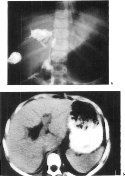
Hepatic artery thrombosis (HAT) is a dreadful complication of orthotopic liver transplantation (OLT). This complication occurred in 27 grafts (68% = 27/393 grafts) in 25 patients (9% = 25/313 patients). HAT was responsible for a high mortality (64% = 16/25 patients) despite a high retransplantation rate (70% = 19/27 grafts). HAT should be suspected in case of fulminant liver failure, delayed bile leak or unexplained fever of sepsis of unknown etiology occurring after liver transplantation. Pulsed doppler examination and arteriogram are the decisive diagnostic procedures. Patients presenting HAT can only be rescued by early diagnosis and retransplantation. Aneurysms of the hepatic arterial supply must also be treated urgently, either by conventional vascular repair if possible or by retransplantation, because or the high incidence of fatal rupture (3/4 patients = 75%).


Figures

Fig. 1
Fig. 1
Intra-operative view and angiography of hepatic allograft revascularization using the fold-over technique in case of triple arterial allograft supply. *SMA with right HA (RHA); °CA with middle (MHA) and left HA (LHA).
Fig. 2
Fig. 2
Intra-operative view and angiography of iliac vessel graft. This graft is anastomosed to the donor hepatic artery (↓), passed behind the neck of the pancreas (*) and anastomosed to the infrarenal abdominal aorta of recipient. PV = portal vein; BD = donor bile duct.
Fig. 3
Fig. 3
Angiography and CT-scan: showing massive gangrene of the allograft due to HAT; one can see the gazbubbles in the right hypochonder.
Fig. 4
Fig. 4
Percutaneous transcutaneous cholangiography (PTC) and CT-scan: showing intrahepatic `biloma' formation secondary to HAT.
Fig. 5
Fig. 5
PTe: biliary stricture of choledochojejunostomy secondary to HAT. The angiography shows a well collateralized allograft.
Fig. 6
Fig. 6
Pseudoaneurysm at the anastomosis between donor and recipient aorta (↓) on angiography and CT-scan.
Fig. 7
Fig. 7
Limited liver allograft infarction (*) due to embolization (↓) of the posterolateral arterial branch of the graft.

References

    1. Gordon RD, Shaw BW, jr., Iwatsuki S, Starzl TE. A simplified technique for revascularization of liver homografts with a variant right hepatic artery from the superior mesenteric artery. Surg. Gynec. Obstet. 1985;160:474–476. - PMC - PubMed
    1. Gordon RD, Makowka L, Bronsther MO, Lerut J, Esquivel CO, Iwatsuki S, Starzl TE. Complications of liver transplantation. In: Toledo-Peyrera LH, editor. Complications of organ transplantation. Marcel Dekker Inc.; New York: 1987. pp. 329–354.
    1. Klintmalm GB, Olson LM, Paulsen AW, Whitten CW, Husberg B. Hepatic artery thrombosis after liver transplantation, electromagnetic blood flow evaluation. Transplant. Proc. 1988 in press. - PubMed
    1. Lerut J, Gordon RD, Iwatsuki S, Shaw BW, jr., Esquivel CO, Tzakis A, Starzl TE. Biliary tract complications in 393 human liver transplantations. Transplantation. 1987;43:47–51. - PMC - PubMed
    1. Segal MC, Zajko AB, Bowen A, Skolnick ML, Bron KM, Penkrot RJ, Slasky BS, Starzl TE. Doppler ultrasound as a screen for hepatic artery thrombosis after liver transplantation. Transplantation. 1986;41:539–541. - PMC - PubMed

LinkOut - more resources