Review
. 2019 Feb;157(2):R33-R51.
doi: 10.1530/REP-18-0350.

It takes two (centrioles) to tango

Affiliations
Review

It takes two (centrioles) to tango

Tomer Avidor-Reiss et al. Reproduction. 2019 Feb.

Abstract

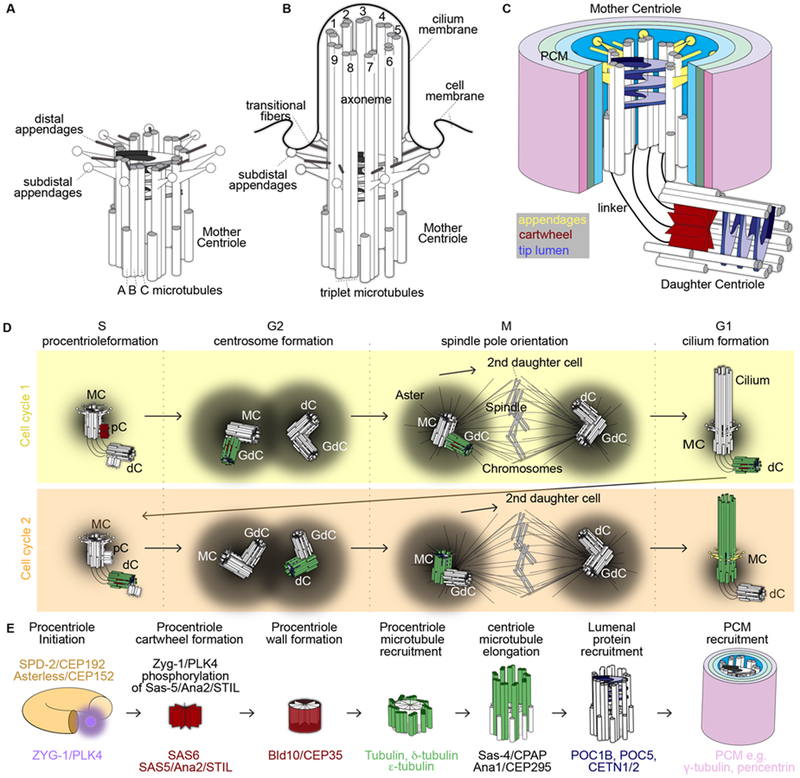
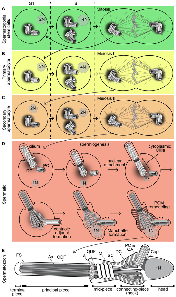
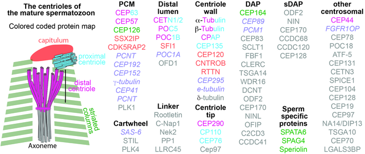
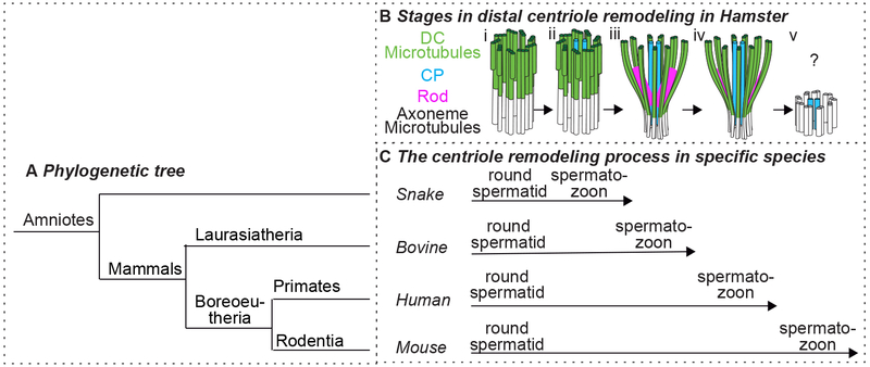
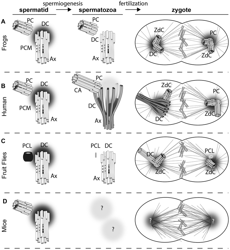
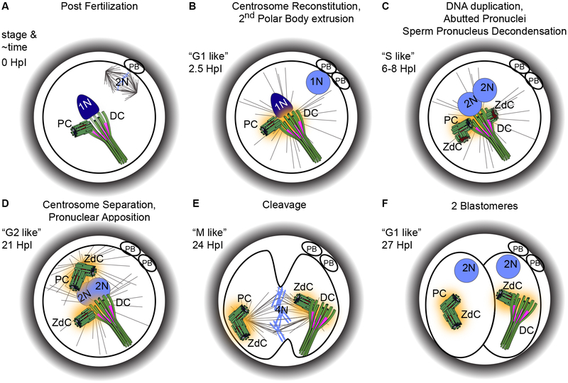
Cells that divide during embryo development require precisely two centrioles during interphase and four centrioles during mitosis. This precise number is maintained by allowing each centriole to nucleate only one centriole per cell cycle (i.e. centriole duplication). Yet, how the first cell of the embryo, the zygote, obtains two centrioles has remained a mystery in most mammals and insects. The mystery arose because the female gamete (oocyte) is thought to have no functional centrioles and the male gamete (spermatozoon) is thought to have only one functional centriole, resulting in a zygote with a single centriole. However, recent studies in fruit flies, beetles and mammals, including humans, suggest an alternative explanation: spermatozoa have a typical centriole and an atypical centriole. The sperm typical centriole has a normal structure but distinct protein composition, whereas the sperm atypical centriole is distinct in both. During fertilization, the atypical centriole is released into the zygote, nucleates a new centriole and participates in spindle pole formation. Thus, the spermatozoa's atypical centriole acts as a second centriole in the zygote. Here, we review centriole biology in general and especially in reproduction, we describe the discovery of the spermatozoon atypical centriole, and we provide an updated model for centriole inherence during sexual reproduction. While we focus on humans and other non-rodent mammals, we also provide a broader evolutionary perspective.


Conflict of interest statement

Declarations

The authors declare that they do not have any conflicts of interest.

Figures

Figure 1
Figure 1. Typical centriole structure and function in a dividing cell.
A-C) Structure of the Mother Centriole (MC) (A), when it extends to form a cilium (B), and as part of a centrosome with a Daughter Centriole (dC) (C). D) The centriole through the cell cycle. A cell has four centrioles in S, G2, and M phases of the cell cycle and two centrioles in G1. It takes multiple steps throughout the two cell cycles for a centriole to mature into a mother; centriole initiation begins as a procentriole (pC) that then develops into a granddaughter centriole (GdC), then to a dC, and finally matures into a MC. The figure depicts the centriole’s initiation and maturation with a single centriole highlighted in color in relation to the other centriole in grayscale. Red – cartwheel, Green- microtubules, Yellow – appendages. E) Centriole initiation and maturation with specific attention to proteins and their functions.
Figure 2
Figure 2. The centriole during human spermatogenesis and in the spermatozoon.
The formation and function of a centriole during the differentiation of a spermatogonial stem cell (A), primary spermatocyte (B), secondary spermatocyte (C), spermatid (D), to a spermatozoon (E). 1N, 2N, and 4N: number of chromosome ploidy in the nucleus; G1, S, M: cell cycle phases. The pictures are not to scale. Ax, axoneme; FS, Fibrous Sheath; A, annulus; M, mitochondrion; ODF, Outer Dense Fibers; SC, Striated Column; Cap, Capitulum; CA; Centriolar Adjunct; PC, Proximal Centriole; DC, Distal Centriole.
Figure 3
Figure 3. Map of Centrosome Proteins in the Spermatozoa Proteins are grouped based on their location in a typical centriole (Linker, Cartwheel, Centriole wall, etcetera).
Centriole protein names are color-coded based on their location in the sperm remodeled centriole (capitulum, striated columns, Proximal Centriole (PC), Distal Centriole (DC), and axoneme). Purple italic font marks proteins that were investigated, but are absent from the remodeled centriole. Gray font marks proteins that were not tested in the spermatozoon. Bi-color proteins are present in 2 locations corresponding to the color schema in the sperm centriole drawing.
Figure 4:
Figure 4:. Various animal species exhibit some or all steps in DC remodeling
A) Phylogenetic tree depicting the relative evolutionary position of snake, bovine, human, and mouse. B) Stages in DC remodeling during spermiogenesis of hamster. The centriole is penetrated by the central pair (CP), then has splayed microtubules, then robust rods appears, then they reduce in size, and finally, the microtubules and rods disappear. Whether there is a very atypical DC structure or no DC at all at the end of the remodeling is unknown. C) The associated stages of DC remodeling in spermiogenesis of four animal species. the DC is progressively more modified in snake, bovine, human, and mouse. The DC of snake spermatozoon is penetrated by the CP (Bii). The DC of bovine spermatozoa are penetrated by the CP, and also has splayed microtubules and robust rods (Biii). The DC of human spermatozoon is penetrated by the center pair, has splayed microtubules, and the rods are present, but are of reduced size (Biv). The mouse appears to have no DC (Bv).
Figure 5
Figure 5. Models of centriole inheritance in various animal species.
Model of sperm centriole inheritance in (A) Frogs, (B) Human, (C) Flies, and (D) Mice. Proximal centriole, PC; Distal Centriole, DC; Zygotic daughter Centriole, ZdC; Axoneme, Ax; Proximal Centriole-Like structure, PCL; Ca, centriole adjunct.
Figure 6
Figure 6. The centrioles in the zygote
When the sperm fuses with the oocyte, the oocyte is arrested at Meiosis II (A). Upon entry, the oocyte finishes the second meiotic division and extrudes the second polar body, while the sperm centrosome (DC and PC) is reconstituted and forms a large aster (B). Then the maternal pronucleus migrates towards the paternal pronucleus and the centrioles begin duplication (producing two zygotic daughter centrioles), presumably while the DNA is duplicating (C). Next, the zygote centrosomes separate from each other while the pronuclei begin to merge (D). Then the zygote undergoes mitosis, much like a normal dividing cell (E), and this division results in two blastomeres (F), which will not form cilia like a normal cell in G0/G1. These two blastomeres will alternate between M and S phases; they will not form cilia or exhibit cytoplasmic growth until the blastocyst stage (Artus et al. 2006). The timing of the events in approximate Hours post Insemination (HpI) and the similar phase (stage) in regular cell cycles are indicated and timing approximations are based on (Mio & Maeda 2008). The exact timing of centriole duplication is unknown; this drawing is based on parsimony and assumptions based on normal centriole duplication. PB, Polar Body; 1N, 2N, 4N, chromosome ploidy; DC, Distal Centriole; PC, Proximal Centriole; ZdC, Zygotic daughter Centriole.

References

    1. Arquint C & Nigg EA 2016. The PLK4-STIL-SAS-6 module at the core of centriole duplication. Biochem Soc Trans 44 1253–1263. - PMC - PubMed
    1. Artus J, Babinet C & Cohen-Tannoudji M 2006. The cell cycle of early mammalian embryos: lessons from genetic mouse models. Cell Cycle 5 499–502. - PubMed
    1. Avidor-Reiss T 2018. Rapid Evolution of Sperm Produces Diverse Centriole Structures that Reveal the Most Rudimentary Structure Needed for Function. Cells 7 67. - PMC - PubMed
    1. Avidor-Reiss T, Ha A & Basiri ML 2017. Transition Zone Migration: A Mechanism for Cytoplasmic Ciliogenesis and Postaxonemal Centriole Elongation. Cold Spring Harb Perspect Biol. - PMC - PubMed
    1. Avidor-Reiss T & Leroux MR 2015. Shared and Distinct Mechanisms of Compartmentalized and Cytosolic Ciliogenesis. Curr Biol 25 R1143–1150. - PMC - PubMed

Publication types

LinkOut - more resources