. 2018 Nov 29;16(11):e2006577.
doi: 10.1371/journal.pbio.2006577. eCollection 2018 Nov.

Extensive editing of cellular and viral double-stranded RNA structures accounts for innate immunity suppression and the proviral activity of ADAR1p150

Affiliations

Extensive editing of cellular and viral double-stranded RNA structures accounts for innate immunity suppression and the proviral activity of ADAR1p150

Christian K Pfaller et al. PLoS Biol. .

Abstract

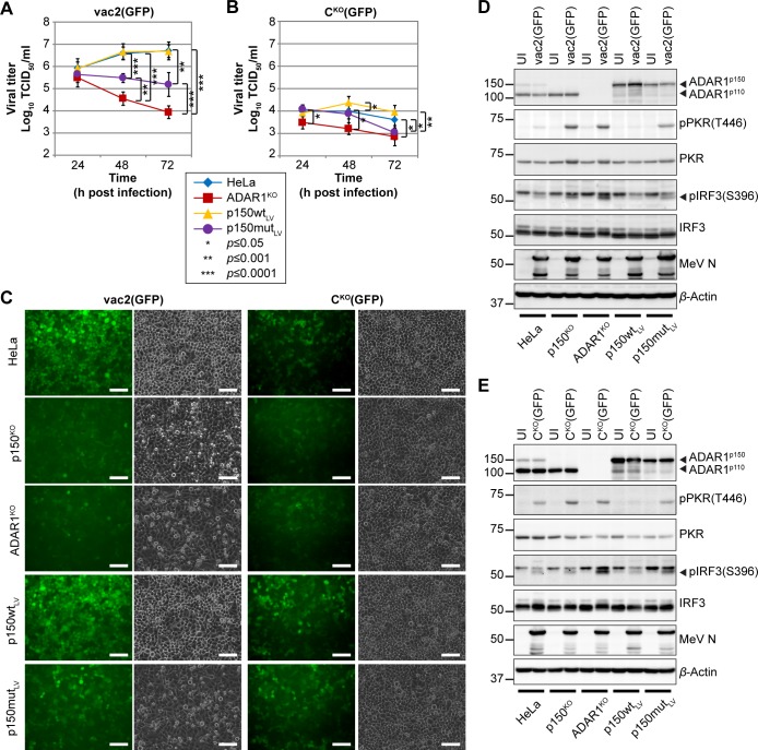
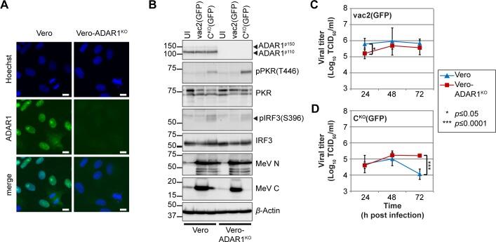
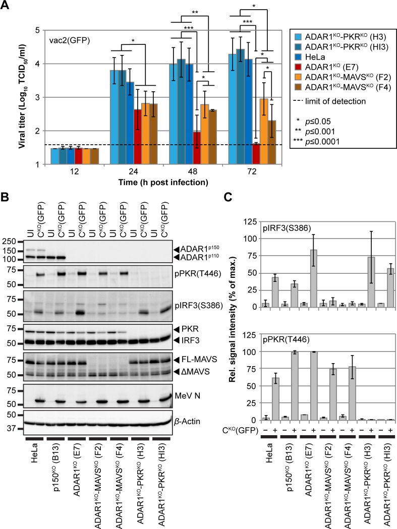
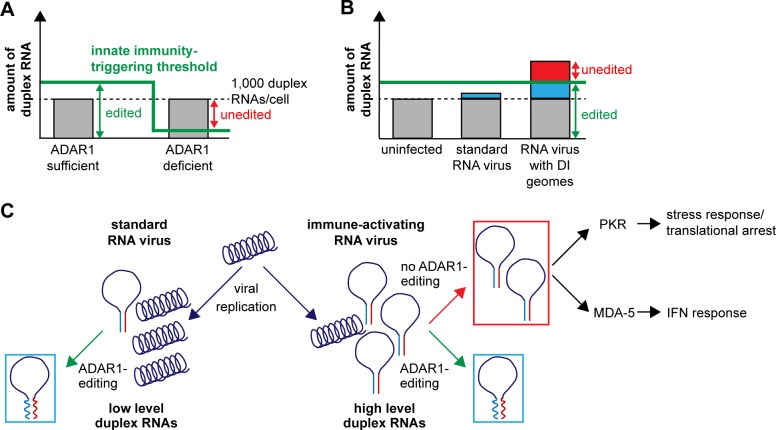
The interferon (IFN)-mediated innate immune response is the first line of defense against viruses. However, an IFN-stimulated gene, the adenosine deaminase acting on RNA 1 (ADAR1), favors the replication of several viruses. ADAR1 binds double-stranded RNA and converts adenosine to inosine by deamination. This form of editing makes duplex RNA unstable, thereby preventing IFN induction. To better understand how ADAR1 works at the cellular level, we generated cell lines that express exclusively either the IFN-inducible, cytoplasmic isoform ADAR1p150, the constitutively expressed nuclear isoform ADAR1p110, or no isoform. By comparing the transcriptome of these cell lines, we identified more than 150 polymerase II transcripts that are extensively edited, and we attributed most editing events to ADAR1p150. Editing is focused on inverted transposable elements, located mainly within introns and untranslated regions, and predicted to form duplex RNA structures. Editing of these elements occurs also in primary human samples, and there is evidence for cross-species evolutionary conservation of editing patterns in primates and, to a lesser extent, in rodents. Whereas ADAR1p150 rarely edits tightly encapsidated standard measles virus (MeV) genomes, it efficiently edits genomes with inverted repeats accidentally generated by a mutant MeV. We also show that immune activation occurs in fully ADAR1-deficient (ADAR1KO) cells, restricting virus growth, and that complementation of these cells with ADAR1p150 rescues virus growth and suppresses innate immunity activation. Finally, by knocking out either protein kinase R (PKR) or mitochondrial antiviral signaling protein (MAVS)-another protein controlling the response to duplex RNA-in ADAR1KO cells, we show that PKR activation elicits a stronger antiviral response. Thus, ADAR1 prevents innate immunity activation by cellular transcripts that include extensive duplex RNA structures. The trade-off is that viruses take advantage of ADAR1 to elude innate immunity control.


Conflict of interest statement

The authors have declared that no competing interests exist.

Figures

Fig 1
Fig 1. ADAR1 edits cellular transcripts forming duplex RNA structures.
(A) Western blot analysis of untreated and IFN A/D–treated ADAR1-modified cells. Cells were treated with 1,000 U/ml IFN A/D for 24 h. (B) Ratios of absolute numbers of identified variants in HeLa and ADAR1KO cells. A>G and U>C variants show higher enrichment in HeLa cells. Average and standard deviation of numbers of variants are indicated. P values of each variant were determined by one-sample, two-tailed Student’s t test against an expected value of 1 (***, P ≤ 0.0001). Underlying values can be found in S1 Data. (C) Relative contribution of ADAR1 isoforms (p150 and p110) to editing in the top 156 genes and correlation with editing in exons/UTRs or introns. Values are calculated based on the number of editing sites detected by GIREMI in HeLa, p150KO, and ADAR1KO cells. Non-ADAR1, percentage of editing sites remaining in ADAR1KO cells. (D) Coverage plot of the 3′ UTR region of VOPP1. The region in the red box shows strong enrichment of U>C transitions. Nucleotide positions with variant frequencies ≥10% are color-coded: A, green; C, blue; G, orange; U, red. (E) Editing score analysis of the boxed region of (D) in HeLa, p150KO, ADAR1KO, and HeLa cells treated with 1,000 U/ml IFN A/D (top to bottom). Gray line indicates total coverage (“Cov.”) in the region. Repetitive sequences are indicated at the bottom: positive sense in blue and negative sense in red. (F) Correlation of editing scores of the VOPP1-region in HeLa + IFN A/D against untreated HeLa cells (gray dots, black line), HeLa versus p150KO (red dots and line), and HeLa versus ADAR1KO (blue dots and line). (G) Relative editing rates in cell lines and primary RNAseq data. Editing rates are normalized to coverage and length of the analyzed regions. Editing rate of each gene in HeLa cells is set to 100%. ADAR1, adenosine deaminase acting on RNA 1; ADAR1KO, fully ADAR1-deficient; GIREMI, Genome-independent Identification of RNA Editing by Mutual Information; IFN A/D, recombinant type-I interferon-alpha; lin. regr., linear regression; N/A, no RNAseq data available because of low or no coverage; p150KO, selectively ADAR1p150-deficient; p150mutLV, catalytically inactive ADAR1p150; p150wtLV, wild-type ADAR1p150; PKR, protein kinase R; pPKR, phospho-PKR; RNAseq, RNA sequencing; UTR, untranslated region; VOPP1, vesicular, overexpressed in cancer, prosurvival protein 1.
Fig 2
Fig 2. ADAR1-editing patterns are conserved in different tissues of primates.
(A) Editing scores in the 3′ UTR region of NDUFS1 (antisense gene) in HeLa, p150KO, and ADAR1KO cells (from top to bottom). (B) Organization of transposable elements in the 3′ UTR of NDUFS1. Positive-sense elements are shown in blue, negative-sense elements in red. Top half shows the human gene (H.sa.), bottom half the macaque (M.mul.) gene. Yellow highlighted regions are conserved across species. Green numbers and letters refer to approximate positions in secondary structures in S6A Fig and S6B Fig. (C) Editing scores in the 3′ UTR region of macaque NDUFS1 derived from RNAseq data of cerebellum and spleen [39]. ADAR1, adenosine deaminase acting on RNA 1; ADAR1KO, fully ADAR1-deficient; H.sa., Homo sapiens; M.mul., M. mulatta; NDUFS1, NADH:ubiquinone oxidoreductase core subunit S1; p150KO, selectively ADAR1p150-deficient; RNAseq, RNA sequencing; UTR, untranslated region.
Fig 3
Fig 3. ADAR1 editing in cellular transcripts sets an immune-activation threshold for dsRNA.
(A) Quantification of ADAR1-edited transcripts (order as in S1 Table) in HeLa, p150KO, and ADAR1KO cells. Four conditions were analyzed for each cell type: UI, infected with MeV-vac2(GFP), infected with MeV-CKO(GFP) (both at MOI = 3 for 24 h), or treated with IFN A/D (1,000 U/ml for 24 h). Heatmap shows log10 FPKM values from RNAseq analysis. (B) Normalized expression levels of ADAR1-edited transcripts relative to GAPDH levels. Median levels for the four conditions described in (A) are shown. Error bars indicate 95% confidence interval. (C) Proportion of ADAR1-edited (red) and nonedited (gray) in HeLa, p150KO, and ADAR1KO cells. (D) Coverage plots and location of ADAR1 editing relative to annotated transcript 3′ end (blue dashed line). Editing occurs between red triangles. Black dot indicates CDS stop codon. (E) Comparison of ADAR1-edited transcripts identified by us (green, cyan, yellow, white), Chung and colleagues [34] (blue, cyan, magenta, white), and Ahmad and colleagues [42] (red, magenta, yellow, white). Each transcript is represented by a single tile. The total numbers in each group (unique, shared by two or three independent studies) are indicated in the legend to the lower right. ADAR1, adenosine deaminase acting on RNA 1; ADAR1KO, fully ADAR1-deficient; CDS, coding sequence; dsRNA, double-stranded RNA; FPKM, fragments per kilobase of transcript per million mapped reads; GAPDH, glyceraldehyde 3-phosphate dehydrogenase; IFN A/D, recombinant type-I interferon-alpha; MOI, multiplicity of infection; NT, nucleotide; p150KO, selectively ADAR1p150-deficient; RNAseq, RNA sequencing; UI, uninfected.
Fig 4
Fig 4. ADAR1 edits MeV genomes and is required for efficient viral replication.
(A and B) Growth curve analyses of (A) MeV-vac2(GFP) and (B) MeV-CKO(GFP) in HeLa cell lines infected at an MOI of 0.1 and harvested at the time points indicated. Values are average ± standard deviation of n = 5 for each time point. For p150KO cells, 3 replicates were generated on clone B13 and 2 replicates on clone C10. For ADAR1KO cells, 3 replicates were generated on clone E7 and 2 replicates on clone E2. Significance was determined by unpaired two-tailed Student’s t test and is indicated with asterisks (*, P < 0.05; ***, P < 0.0001). Underlying values can be found in S1 Data. (C) Absolute number of viral reads with >5 mutations (“NM>5”) in RNAseq samples. Underlying values can be found in S1 Data. (D) Frequency of these reads relative to total number of MeV-specific reads. U>C: reads with predominantly U>C mutations (red); A>G: reads with predominantly A>G mutations (green). Underlying values can be found in S1 Data. (E) Editing scores of MeV-vac2(GFP) and MeV-CKO(GFP) genomes from HeLa, p150KO, and ADAR1KO infections. Scores are shown for transitions (A>G, green; U>C, red; G>A, orange; C>U, blue) and a read coverage (gray) of at least 10. (F and G) Correlation of (F) MeV-vac2(GFP) and (G) MeV-CKO(GFP) genome editing between HeLa and p150KO cells (gray dots and black line) or HeLa and ADAR1KO cells (red dots and line). ADAR1, adenosine deaminase acting on RNA 1; ADAR1KO, fully ADAR1-deficient; KO, knock-out; lin. regr., linear regression; MeV, measles virus; MOI, multiplicity of infection; NT, nucleotide; p150KO, selectively ADAR1p150-deficient; RNAseq, RNA sequencing.
Fig 5
Fig 5. ADAR1p150 rescues MeV growth restriction and suppresses intrinsic immunity activation.
(A and B) Growth curve analyses of (A) MeV-vac2(GFP) and (B) MeV-CKO(GFP) in HeLa, ADAR1KO (clone E7), p150wtLV, and p150mutLV cells infected at an MOI of 0.1 and harvested at the time points indicated. Values are average ± standard deviation of n = 6 for each time point. P values were determined by unpaired, two-tailed Student’s t test (*, P < 0.05; **, P < 0.001; ***, P < 0.0001). Underlying values can be found in S1 Data. (C) GFP expression of MeV-vac2(GFP) and MeV-CKO(GFP) in ADAR1-modified cells infected at an MOI of 3. Images were taken at 24 h post infection and show GFP fluorescence (green signal) and corresponding phase contrast. Scale bar equals 100 μm. (D and E) Western blot analyses of MeV-vac2(GFP) (D) and MeV-CKO(GFP) (E) infected cell lysates. Cells were infected at an MOI of 3 or were left UI and harvested 24 h post infection. ADAR1, adenosine deaminase acting on RNA 1; ADAR1KO, fully ADAR1-deficient; GFP, green fluorescent protein; IRF3, interferon regulatory transcription factor 3; MeV, measles virus; MOI, multiplicity of infection; N, nucleoprotein; p150KO, selectively ADAR1p150-deficient; p150mutLV, catalytically inactive ADAR1p150; p150wtLV, wild-type ADAR1p150; pIRF3, phospho-IRF3; PKR, protein kinase R; pPKR, phospho-PKR; UI, uninfected.
Fig 6
Fig 6. ADAR1 only has a minor effect on MeV replication in IFN-incompetent Vero cells.
(A) Confocal immunofluorescence staining of Vero and Vero-ADAR1KO cells. Nuclear staining (Hoechst) in blue, ADAR1-specific staining in green. Scale bar equals 10 μm. (B) Western blot analysis of Vero and Vero-ADAR1KO cell lysates UI or infected with MeV-vac2(GFP) or MeV-CKO(GFP) at an MOI of 0.1, 32 h post infection. (C and D) Growth curve analyses of (C) MeV-vac2(GFP) and (D) MeV-CKO(GFP) in Vero and Vero-ADAR1KO cells infected at an MOI of 0.1 and harvested at indicated time points. Values are average ± standard deviation of n = 6 for each time point. P values were determined by unpaired, two-tailed Student’s t test (*, P ≤ 0.05; ***, P ≤ 0.0001). Underlying values can be found in S1 Data. ADAR1, adenosine deaminase acting on RNA 1; ADAR1KO, fully ADAR1-deficient; IFN, interferon; IRF3, IFN regulatory transcription factor 3; MeV, measles virus; MOI, multiplicity of infection; N, nucleoprotein; pIRF3, phospho-IRF3; PKR, protein kinase R; pPKR, phospho-PKR; UI, uninfected.
Fig 7
Fig 7. PKR activation elicits a stronger antiviral response than MAVS-mediated IFN induction.
(A) Growth curve analyses of MeV-vac2(GFP) in HeLa, ADAR1KO (clone E7), ADAR1KO-MAVSKO (clones F2 and F4), and ADAR1KO-PKRKO cells (clones H3 and HI3) infected at an MOI of 0.1 and harvested at indicated time points. Values are average ± standard deviation of n = 6 for each time point. P values were determined by unpaired, two-tailed Student’s t test (*, P ≤ 0.05; **, P ≤ 0.001; ***, P ≤ 0.0001). Underlying values can be found in S1 Data. (B) Western blot analysis of cell lysates of UI or MeV-CKO(GFP)-infected cells at an MOI of 3, 24 h post infection. (C) Quantification of the pIRF3(S386) signals (top panel) and pPKR(T446) signals (bottom panel) shown in (B). Values are average ± standard deviation of two independent experiments. Underlying values can be found in S1 Data. ADAR1, adenosine deaminase acting on RNA 1; ADAR1KO, fully ADAR1-deficient; FL-MAVS, full-length MAVS; IFN, interferon; IRF3, IFN regulatory transcription factor 3; MAVS, mitochondrial antiviral signaling protein; MeV, measles virus; MOI, multiplicity of infection; N, nucleoprotein; p150KO, selectively ADAR1p150-deficient; pIRF3, phospho-IRF3; PKR, protein kinase R; pPKR, phospho-PKR; UI, uninfected.
Fig 8
Fig 8. Models for the regulatory function of ADAR1 in autoimmunity and infection.
(A) In ADAR1-sufficient cells, transcribed cellular duplex RNA (about 1,000 transcripts under homeostatic conditions, black dashed line) is efficiently edited (green arrow). Thus, no activation of innate immunity occurs. ADAR1 expression level sets the threshold for innate immunity activation (green line). In ADAR1-deficient cells, the threshold is decreased. Levels of transcribed duplex RNA remain equal, but duplexes are not edited and innate immunity is triggered (red arrow). (B) A standard RNA virus (e.g., MeV-vac2) generates low amounts of dsRNA (blue), which is efficiently edited by ADAR1 and thus insufficient to activate innate immunity. In contrast, an RNA virus with DI genomes (e.g., MeV-CKO) generates large amounts of duplex RNA (blue and red). ADAR1 still edits some of it (blue), but unedited dsRNA activates innate immunity (red). (C) Schematic representation of the generation of immunogenic duplex RNA (panhandle structures) during viral infection and the impact of ADAR1 on PKR- and MDA-5-mediated innate immunity activation by these RNAs. ADAR1, adenosine deaminase acting on RNA 1; DI, defective interfering; dsRNA, double-stranded RNA; IFN, interferon; MDA-5, melanoma differentiation–associated gene 5; MeV-CKO, MeV unable to express C protein; PKR, protein kinase R.

Similar articles

Cited by

References

    1. Schneider WM, Chevillotte MD, Rice CM. Interferon-stimulated genes: a complex web of host defenses. Annu Rev Immunol. 2014;32:513–45. 10.1146/annurev-immunol-032713-120231 - DOI - PMC - PubMed
    1. Hartner JC, Walkley CR, Lu J, Orkin SH. ADAR1 is essential for the maintenance of hematopoiesis and suppression of interferon signaling. Nat Immunol. 2009;10(1):109–15. 10.1038/ni.1680 - DOI - PMC - PubMed
    1. Bass BL, Weintraub H. An unwinding activity that covalently modifies its double-stranded RNA substrate. Cell. 1988;55(6):1089–98. 10.1016/0092-8674(88)90253-X - DOI - PubMed
    1. Wagner RW, Smith JE, Cooperman BS, Nishikura K. A double-stranded RNA unwinding activity introduces structural alterations by means of adenosine to inosine conversions in mammalian cells and Xenopus eggs. Proc Natl Acad Sci U S A. 1989;86(8):2647–51. 10.1073/pnas.86.8.2647 - DOI - PMC - PubMed
    1. Nishikura K. A-to-I editing of coding and non-coding RNAs by ADARs. Nat Rev Mol Cell Biol. 2016;17(2):83–96. 10.1038/nrm.2015.4 - DOI - PMC - PubMed

Publication types

MeSH terms