Review
. 2019 Apr 1;40(2):369-416.
doi: 10.1210/er.2018-00075.

Ovulation: Parallels With Inflammatory Processes

Affiliations
Review

Ovulation: Parallels With Inflammatory Processes

Diane M Duffy et al. Endocr Rev. .

Abstract

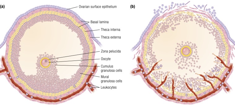
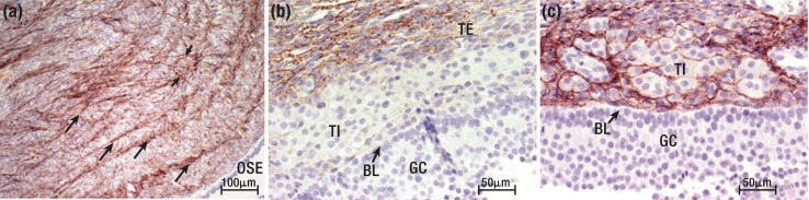
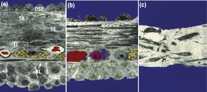
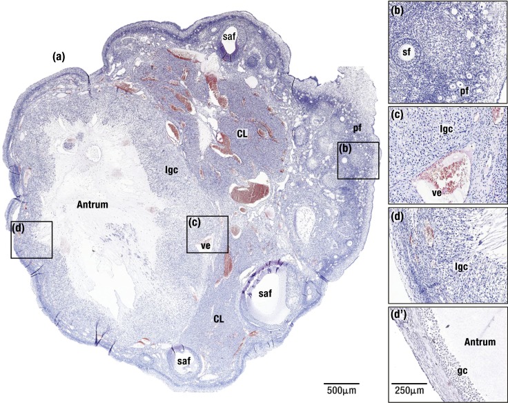
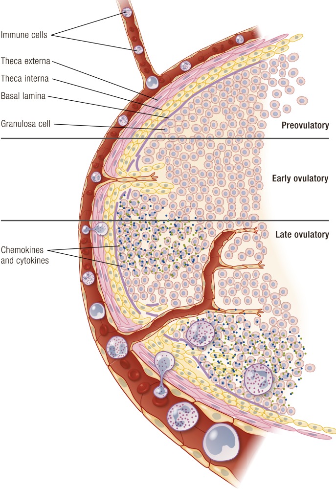
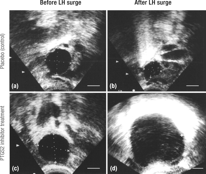
The midcycle surge of LH sets in motion interconnected networks of signaling cascades to bring about rupture of the follicle and release of the oocyte during ovulation. Many mediators of these LH-induced signaling cascades are associated with inflammation, leading to the postulate that ovulation is similar to an inflammatory response. First responders to the LH surge are granulosa and theca cells, which produce steroids, prostaglandins, chemokines, and cytokines, which are also mediators of inflammatory processes. These mediators, in turn, activate both nonimmune ovarian cells as well as resident immune cells within the ovary; additional immune cells are also attracted to the ovary. Collectively, these cells regulate proteolytic pathways to reorganize the follicular stroma, disrupt the granulosa cell basal lamina, and facilitate invasion of vascular endothelial cells. LH-induced mediators initiate cumulus expansion and cumulus oocyte complex detachment, whereas the follicular apex undergoes extensive extracellular matrix remodeling and a loss of the surface epithelium. The remainder of the follicle undergoes rapid angiogenesis and functional differentiation of granulosa and theca cells. Ultimately, these functional and structural changes culminate in follicular rupture and oocyte release. Throughout the ovulatory process, the importance of inflammatory responses is highlighted by the commonalities and similarities between many of these events associated with ovulation and inflammation. However, ovulation includes processes that are distinct from inflammation, such as regulation of steroid action, oocyte maturation, and the eventual release of the oocyte. This review focuses on the commonalities between inflammatory responses and the process of ovulation.


Figures

Figure 1.
Figure 1.
The preovulatory follicle. (a) Preovulatory follicle prior to the LH surge. The oocyte is surrounded by the zona pelucida and cumulus granulosa cells that connect to the mural granulosa cells that line the interior of the follicle. The granulosa cell compartment is separated from the theca cell compartment by a basal lamina. The theca cell compartment is composed of an inner theca interna and an outer theca externa. Unlike the granulosa cell compartment, the theca cell layer is highly vascularized (red). Circulating leukocytes are present in the vessels. The theca externa blends into a layer of connective tissue that is separated from the ovarian surface epithelium by a basal lamina. (b) Preovulatory follicle following LH stimulation immediately prior to ovulation. Disruption of the granulosa cell basal lamina allows extension of vessels into the granulosa cell compartment. Theca cells and leukocytes also enter into the granulosa cell compartment. The cumulus oocyte complex detaches from the surrounding granulosa cells and undergoes cumulus expansion. At the follicular apex (top of image), there is a loss of ovarian surface epithelium, the breakdown of the underlying basal lamina, and a loss of theca cells and granulosa cells. Rupture will occur at the follicle apex.
Figure 2.
Figure 2.
Collagens in the human ovary and ovulatory follicle. (a) Collagen type I (brown) in the human ovarian capsular stroma, showing a distribution of collagen type I in concentric layers (long arrows) with bundles (short arrows) joining the concentric layers. (b) Collagen type I (brown) in the theca externa (TE) and (c) collagen type III (brown) in the theca interna (TI) of a human preovulatory follicle. Nova Red stain. BL, basal lamina; GC, granulosa cells. [Reproduced with permission from Lind A-K, Weijdegard B, Dahm-Kahler P. Collagens in the human ovary and their changes in the perifollicular stroma during ovulation. Acta Obstet Gynecol Scand 2006;85(12):1476–1484.]
Figure 3.
Figure 3.
Structure of the apex of the rabbit preovulatory follicle. Faux-colored electron microscopic images were obtained (a) before the LH surge, (b) 1 to 2 h prior to follicle rupture, and (c) immediately before follicular rupture. (a) Layers of the intact follicular wall. At the apex is a single layer of ovarian surface epithelium (OSE) containing granules with unknown contents (red). Underlying the OSE is the tunica albuginea (TA) and theca externa (TE), with numerous cells and extracellular connective tissue. Capillaries with red blood cells (red) and steroidogenic theca interna cells (TI, containing yellow lipid droplets) are adjacent to the granulosa cell (GC) basal lamina (BL). (b) Changes in the follicular wall following an LH stimulus. Notable changes include loss of many of the OSE, elongation of fibroblasts and thinning of the ECM in the TA and TI, and fewer granulosa cells. Capillaries contain clotted red blood cells (red), platelets (blue), and immune cells (pink). Granulosa cells now contain many lipid droplets (green), consistent with increased steroid hormone synthesis. In (c), which depicts the follicular apex immediately prior to ovulation, no OSE or granulosa cells remain at the apex. Remaining connective tissue is thin and disorganized. [Color micrographs courtesy of Dr. Lawrence Espey.]
Figure 4.
Figure 4.
(a) Macaque ovary soon after ovulation. On the left, a recently ovulated follicle (∼8 h after follicle rupture) is seen with the antrum surrounded by luteinizing granulosa cells (lgc). Remainder of the ovary contains two regressing corpora lutea (CL) and several small antral follicles (saf). (b) An enlarged view of the ovarian cortex shows numerous primordial follicles (pf) and a secondary follicle (sf). (c and d) The luteinizing granulosa cell layer (lgc) thickens as cells undergo hypertrophy at both the follicle base (c) and apex (d). In (c), an enlarged vessel (ve) is adjacent to the luteinizing granulosa cell layer. (d′) An example of the apical region of a macaque preovulatory follicle shows the thin cortical stroma and few layers of compact granulosa cells (gc) present prior to the LH surge. Hematoxylin and eosin stain.
Figure 5.
Figure 5.
Regional expression of proteases and protease inhibitors. Gelatinase activity (intense green) is localized at the rat follicle apex as ovulation approaches. (a–c) Gelatinase activity predominates in the theca (a) before hCG administration (arrows), (b) in the apical region of the follicle 12 h after hCG (arrow), and (c) throughout the forming corpus luteum (CL). (d) The PA inhibitor PAI-1 (now known as SERPINE1) protein is lower at the follicle apex than at the follicle base (nonapex) just before ovulation in monkey ovulatory follicles; (e) SERPINE1 protein correlates with higher expression of the PGE2 receptor PTGER1 (green) in granulosa cells (gc) at the follicle base when compared with (f) the apex. an, antrum; st, stroma. (a–c) Gelatinase activity visualized with green fluorescence; (e and f) Alexa Fluor 488. [Panels (a)–(c) adapted with permission from Curry TE Jr, Song L, Wheeler SE. Cellular localization of gelatinases and tissue inhibitors of metalloproteinases during follicular growth, ovulation, and early luteal formation in the rat. Biol Reprod 2001;65(3):855–865. Illustration presentation copyright by the Endocrine Society. Panels (d)–(f) adapted with permission from Harris SM, Aschenbach LC, Skinner SM, et al. Prostaglandin E2 receptors are differentially expressed in subpopulations of granulosa cells from primate periovulatory follicles. Biol Reprod 2011;85(5):916–923. Illustration presentation copyright by the Endocrine Society.]
Figure 6.
Figure 6.
Signaling pathways activated by the LH surge in ovulatory follicles. LH activates multiple signaling pathways, including protein kinase A (PKA), protein kinase C (PKC), phosphatidylinositol 3-kinase (PI3K), and p38MAPK. LH also activates the epidermal growth factor receptor (EGFR) signaling pathway through the rapid induction of EGF-like factor expression and shedding of existing EGF-like factors from the membrane. Bold arrows emphasize the importance of PKA and EGFR signaling pathways in granulosa cells of ovulatory follicles. AC, adenylyl cyclase; AKT, Akt/protein kinase B; CREB, cAMP response element binding protein; DAG, 1,2-diacylglycerol; IP3, inositol 1,4,5-triphosphate; MEK, mitogen-activated protein kinase kinase; MMP, matrix metalloproteinase; PIP2, phosphatidylinositol 4,5-bisphosphate; PIP3, phosphatidylinositol 3,4,5-trisphosphate; PLC, phospholipase C.
Figure 7.
Figure 7.
Granulosa and theca cells cooperate to produce steroid hormones. Before the LH surge, (i) theca cells produce predominantly androgens in response to LH, (ii) androgens diffuse to granulosa cells, and (iii) granulosa cells convert androgens to estrogens in response to FSH. After the LH surge, (i) decreased CYP17A1 expression increases progesterone synthesis and decreases androgen synthesis in theca cells, and (ii) increased HSD3B1 increases progesterone synthesis and declining CYP19A1 decreases estrogen synthesis by granulosa cells. The LH surge also increases HSD11B1 and decreases HSD11B2 in granulosa cells to increases synthesis of cortisol from circulating cortisone. Enzymes shown in green increase after the LH surge. Enzymes shown in red decrease after the LH surge.
Figure 8.
Figure 8.
PGE2 synthesis, receptors, transport, and metabolism in granulosa cells. Left: PGE2 synthesis enzymes (blue) are associated with membranes of the nuclear envelope (not shown) and endoplasmic reticulum (ER). PLA2G4A cleaves arachidonic acid (AA) from membrane phospholipids. PTGS2 converts AA into PGH2. PTGES converts PGH2 into bioactive PGE2. PGE2 is converted to an inactive metabolite (15-keto-PGE2) by HPGD (purple). Right: PGE2 acts via four PGE2 receptors (PTGER1, PTGER2, PTGER3, and PTGER4, green). Each PTGER couples to a subset of G proteins (purple); most frequently used and major intracellular signals are shown for plasma membrane PTGERs. PTGERs can also be located in the membranes of the ER and nucleus; G proteins also couple with PTGERs in these locations (not shown). On both panels, multiple methods of PGE2 transport across the plasma membrane have been proposed (blue) and are discussed in the text. [Adapted with permission from Duffy DM. Novel contraceptive targets to inhibit ovulation: the prostaglandin E2 pathway. Hum Reprod Update 2015;21(5):652–670. Illustration presentation copyright of the Endocrine Society.]
Figure 9.
Figure 9.
LH-stimulated changes in capillary growth and leukocyte delivery in the ovulatory follicle. Before the LH surge, immune cells are present in the ovarian vasculature (red) and theca interna of the preovulatory follicle (top). In response to the LH surge, capillary growth and leukocyte invasion begin. During the early ovulatory period, vessels in the theca interna are the source for new capillary growth into the granulosa cell layer. New vessels and breakdown of the granulosa cell basal lamina provide points of entry for circulating leukocytes to reach the theca and granulosa cell layers of the follicle (center). Secretion of chemokines and cytokines (granules) by leukocytes and granulosa cells begins (center). By the late ovulatory period, breakdown of the granulosa cell basal lamina continues, extensive capillary networks form, and additional leukocytes are seen in the granulosa cell layer prior to ovulation (bottom). Leukocyte-secreted chemokines and cytokines increase as ovulation approaches.
Figure 10.
Figure 10.
Blood flow in the human preovulatory follicle. (a and b) Color Doppler and ultrasound (US) images of human ovarian follicles (a) before the LH surge and (b) after the LH surge but before ovulation. For each panel, the left side shows ultrasound with Doppler blood velocity (US + Doppler) and right side shows only Doppler blood velocity. Red represents flow toward the transducer. Blue represents flow away from the transducer. Blood flow is concentrated at the follicle base and is less prominent at the follicle apex as ovulation approaches (b). [Reproduced with permission from Brannstrom M, Zackrisson U, Hagstrom HG, et al. Preovulatory changes of blood flow in different regions of the human follicle. Fertil Steril 1998;69(3):435–442.]
Figure 11.
Figure 11.
The MMP and ADAMTS families. A general model of the structural organization of the more common MMPs is presented. The MMPs and ADAMTSs contain a signal peptide, a propeptide domain with a sulfhydryl group that must be cleaved for activation, and a catalytic domain that contains the zinc binding site. The MMPs also contain a hinge region and a hemopexin-like domain. The gelatinases contain a fibronectin type II domain whereas certain other MMPs contain a furin-susceptible site that allows intracellular activation. The transmembrane-type MMPs contain a transmembrane linker to a cytoplasmic domain. The ADAMTSs contain a disintegrin domain along with regions of thrombospondin repeats. MT-MMP, membrane-type MMP. [Adapted with permission from Parks WC, Wilson CL, Lopez-Boado YS. Matrix metalloproteinases as modulators of inflammation and innate immunity. Nat Rev Immunol 2004;4(8):617–629; and under a Creative Commons CC-BY 4.0 license from Noel A, Gutierrez-Fernandez A, Sounni NE. New and paradoxical roles of matrix metalloproteinases in the tumor microenvironment. Front Pharmacol 2012;3:140. Illustration presentation copyright by the Endocrine Society.]
Figure 12.
Figure 12.
The plasmin–PA–MMP cascade. The PAs, PLAT or PLAU, are able to cleave plasminogen to plasmin. Plasmin has numerous actions, including activation of the MMP system to bring about matrix breakdown. Plasminogen activator action is regulated by PA inhibitors (SERPINE1 and SERPINB2) wheras MMP activity is regulated in the extracellular environment by the MMP inhibitors, the TIMPs.
Figure 13.
Figure 13.
PTGS2 inhibitor prevents follicle rupture in women. (a and c) Ultrasound images show the preovulatory follicle antrum (dark circles) before the LH surge. (b) Placebo treatment (control) results in decreased size of the antrum, indicative of follicle rupture. (d) The PTGS2 inhibitor rofecoxib treatment yields a follicle which continued to enlarge without rupture. [Reproduced with permission from Pall M, Friden BE, Brannstrom M. Induction of delayed follicular rupture in the human by the selective COX-2 inhibitor rofecoxib: a randomized double-blind study. Hum Reprod 2001;16(7):1323–1328.]

References

    1. Bukovsky A, Presl J. Ovarian function and the immune system. Med Hypotheses. 1979;5(4):415–436. - PubMed
    1. Espey LL. Ovulation as an inflammatory reaction—a hypothesis. Biol Reprod. 1980;22(1):73–106. - PubMed
    1. Medzhitov R. Origin and physiological roles of inflammation. Nature. 2008;454(7203):428–435. - PubMed
    1. Espey LL. Ultrastructure of the apex of the rabbit graafian follicle during the ovulatory process. Endocrinology. 1967;81(2):267–276. - PubMed
    1. Auersperg N, Wong AS, Choi KC, Kang SK, Leung PC. Ovarian surface epithelium: biology, endocrinology, and pathology. Endocr Rev. 2001;22(2):255–288. - PubMed

Publication types

Substances