. 2019 Feb 20;47(3):1532-1543.
doi: 10.1093/nar/gky1198.

Developmental Xist induction is mediated by enhanced splicing

Affiliations

Developmental Xist induction is mediated by enhanced splicing

Cheryl Stork et al. Nucleic Acids Res. .

Abstract

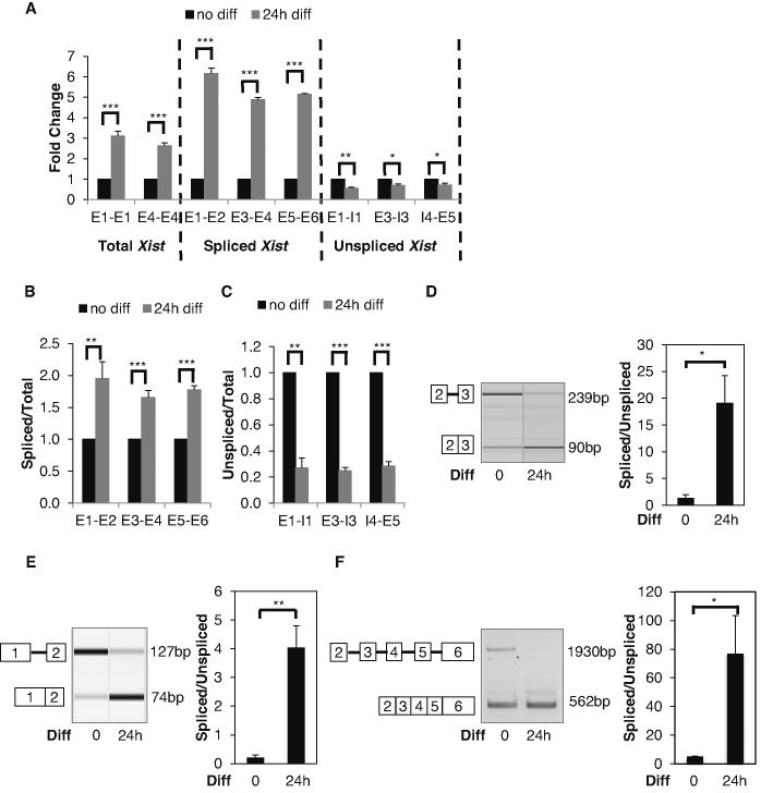
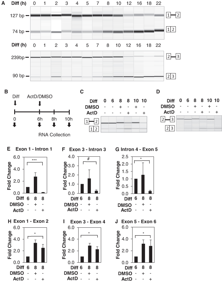
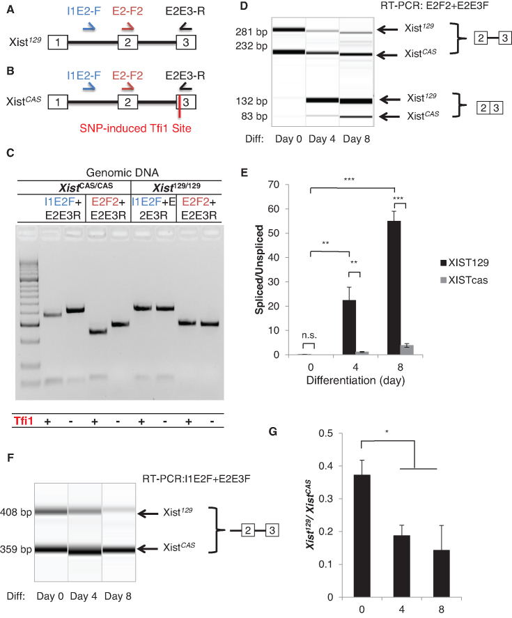
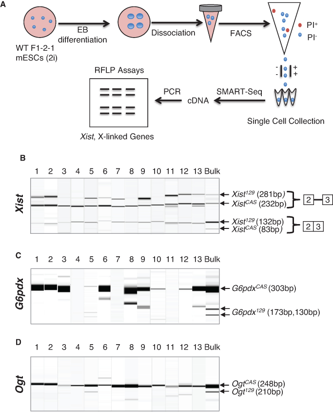
X-inactive-specific transcript (Xist) is a long noncoding RNA (lncRNA) essential for inactivating one of the two X chromosomes in mammalian females. Random X chromosome inactivation is mediated by Xist RNA expressed from the inactive X chromosome. We found that Xist RNA is unspliced in naïve embryonic stem (ES) cells. Upon differentiation, Xist splicing becomes efficient across all exons independent of transcription, suggesting interdependent or coordinated removal of Xist introns. In female cells with mutated polypyrimidine tract binding protein 1 (Ptbp1), differentiation fails to substantially upregulate mature Xist RNA because of a defect in Xist splicing. We further found both Xist129 and XistCAS RNA are unspliced in Mus musculus 129SvJ/Mus castaneous (CAS) hybrid female ES cells. Upon differentiation, Xist129 exhibits a higher splicing efficiency than XistCAS, likely contributing to preferential inhibition of the X129 chromosome. Single cell analysis shows that the allelic choice of Xist splicing is linked to the inactive X chromosome. We conclude post-transcriptional control of Xist RNA splicing is an essential regulatory step of Xist induction. Our studies shed light on the developmental roles of splicing for nuclear-retained Xist lncRNA and suggest inefficient Xist splicing is an additional fail-safe mechanism to prevent Xist activity in ES cells.


Figures

Figure 1.
Figure 1.
Differentiation Induces Xist Splicing. (A) RT-qPCR analysis of total, spliced, and unspliced Xist transcripts. Adherent F1 2-1 cells were differentiated by LIF withdrawal and addition of RA. (B) Ratios of spliced/total Xist transcripts quantified by RT-qPCR. (C) Ratios of unspliced/total Xist transcripts quantified by RT-qPCR. (D) Splicing assay examines intron 2 splicing before and 24 h after differentiation. Representative virtual gel image of intron 2 splicing (left panel). Quantified intron 2 splicing ratio (right panel) (E) Splicing assay examines intron 1 splicing before and 24 h after differentiation. Representative virtual gel image of intron 1 splicing (left panel). Quantified intron 1 splicing ratio (right panel). (F) Splicing assay examines splicing from exon 2 to exon 6 before and 24 h after differentiation. Representative gel image (left panel). PCR cycles were 33 and 29 for undifferentiated and differentiated cells respectively. For differentiated cells, PCR amplification for 33 cycles did not produce the unspliced product and the spliced product was over saturated, so amplification of 29 cycles were used for the presentation. Quantified splicing ratio (right panel). Error bars indicate S.E.M. of three biological replicates. *P-value<0.05; **P-value<0.01; ***P-value<0.001 Student's t-test (one-tailed, unpaired).
Figure 2.
Figure 2.
Enhanced Xist splicing is independent of Xist transcription. (A) Representative RT-PCR virtual gel images of intron 1 splicing (top panel) and intron 2 splicing (bottom panel) in adherent F1 2-1 cells between 0–22 h post differentiation (RA induction and LIF withdrawal). (B) Schematic diagram of experimental procedures. Cells were differentiated for 6 hours before treatment with 5ug/ml Actinomycin D or control DMSO. RNA was collected before differentiation (time 0), for 6 h post differentiation induction (time 6), for 2 and 4 h post treatment (time 8 and time 10). Representative RT-PCR virtual gel image of (C) intron 1 and (D) intron 2splicing following the aforementioned experimental procedures. (E–G) RT-qPCR analysis of unspliced Xist transcripts using primers detecting various introns. (H–J) RT-qPCR of spliced Xist transcripts using primers for various exon–exon junctions. Error bars indicate S.E.M. of three biological replicates. #P-value = 0.06; *P-value < 0.05; ***P-value < 0.001 Student's t-test (one-tailed, unpaired).
Figure 3.
Figure 3.
Allelic difference in Xist splicing is associated with non-random Xist induction. (AB) Schematics of the primer locations and allele-specific restriction enzyme digestion for distinguishing the unspliced and spliced Xist transcripts from the Xist129 and XistCAS alleles. SNP induced Tfi1 cut site is present in exon 3 of the XistCAS allele. (C) A representative gel showing Tfi1 specifically cuts exon 3 of XistCAS/CAS genomic DNA but not Xist129/129 DNA. Genomic DNA was amplified using the I1E2F or the E2F2 forward primer and the E2E3R primer before Tfi1 treatment. (D) Splicing assay detects allele-specific expression of unspliced and spliced transcripts from the Xist129 and XistCAS alleles before and after differentiation. (E) Quantification of intron 2 splicing ratios of XistCAS and Xist129 transcripts, respectively. (F) A representative RT-PCR virtual gel image shows the expression of unspliced XistCAS and unspliced Xist129 transcripts using I1E2-F and E2E3-R primers. (G) Ratios of unspliced Xist129 transcript relative to unspliced XistCAS transcript upon differentiation. Error bars indicate S.E.M. of three biological replicates. ns, P-value > 0.05; *P-value < 0.05; **P-value< 0.01; ***P-value < 0.001.
Figure 4.
Figure 4.
Allele specific Xist splicing is linked to the choice of inactive X chromosome. (A) Schematic diagram of the experimental procedure using embryoid body differentiation, FACS, and SMART-Seq for single cell analysis of allele-specific Xist splicing and allele-specific X-linked gene expression in F1 2-1 cells. (B) Restriction fragment length polymorphism analysis combined with splicing assay detects allelic specific expression of unspliced and spliced Xist transcripts from the X129 and XCAS chromosomes in intermediate cells. (C and D) Restriction fragment length polymorphism analysis detects allelic expression of X-linked genes (C) G6pdx and (D) Ogt from the X129 and XCAS chromosomes. Bulk means RNA samples from bulk cells.

Similar articles

Cited by

References

    1. Huynh K.D., Lee J.T.. Inheritance of a pre-inactivated paternal X chromosome in early mouse embryos. Nature. 2003; 426:857–862. - PubMed
    1. Mak W., Nesterova T.B., de Napoles M., Appanah R., Yamanaka S., Otte A.P., Brockdorff N.. Reactivation of the paternal X chromosome in early mouse embryos. Science. 2004; 303:666–669. - PubMed
    1. Monkhorst K., Jonkers I., Rentmeester E., Grosveld F., Gribnau J.. X inactivation counting and choice is a stochastic process: evidence for involvement of an X-linked activator. Cell. 2008; 132:410–421. - PubMed
    1. Lessing D., Anguera M.C., Lee J.T.. X chromosome inactivation and epigenetic responses to cellular reprogramming. Annu. Rev. Genomics Hum. Genet. 2013; 14:85–110. - PubMed
    1. Augui S., Nora E.P., Heard E.. Regulation of X-chromosome inactivation by the X-inactivation centre. Nat. Rev. Genet. 2011; 12:429–442. - PubMed

Publication types

MeSH terms

Substances